. 2004 Jun;24(11):4880-94.
doi: 10.1128/MCB.24.11.4880-4894.2004.

Amphoterin stimulates myogenesis and counteracts the antimyogenic factors basic fibroblast growth factor and S100B via RAGE binding

Affiliations

Amphoterin stimulates myogenesis and counteracts the antimyogenic factors basic fibroblast growth factor and S100B via RAGE binding

Guglielmo Sorci et al. Mol Cell Biol. 2004 Jun.

Abstract

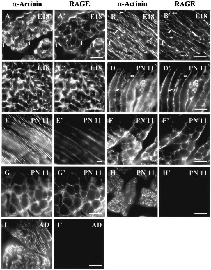
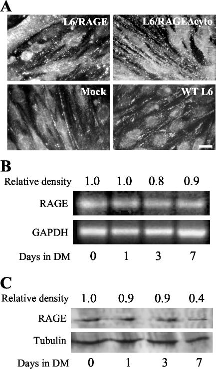
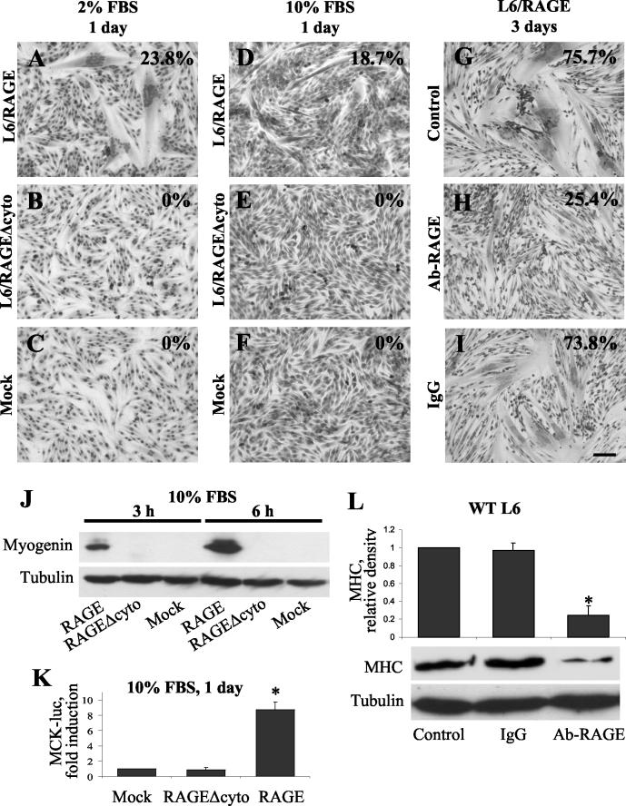
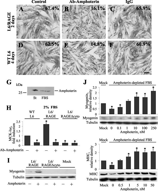
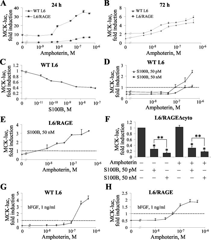
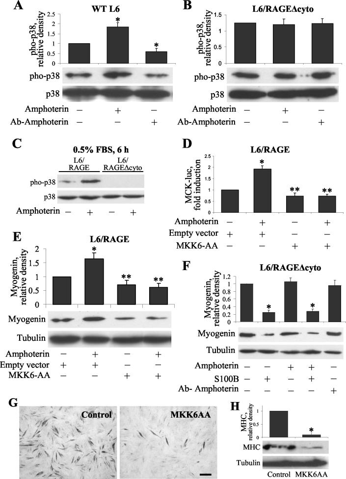
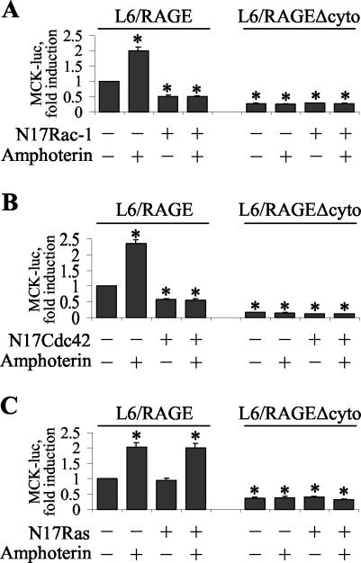
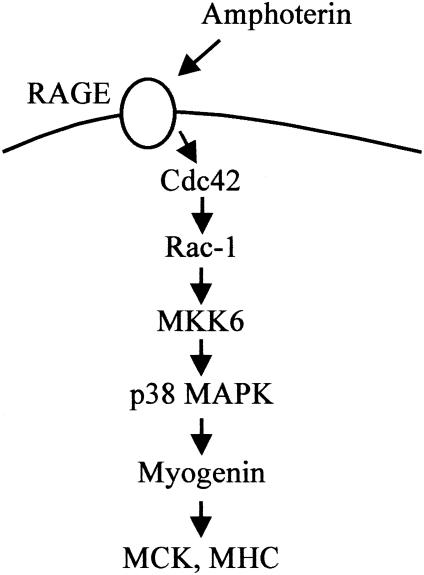
The receptor for advanced glycation end products (RAGE), a multiligand receptor of the immunoglobulin superfamily, has been implicated in the inflammatory response, diabetic angiopathy and neuropathy, neurodegeneration, cell migration, tumor growth, neuroprotection, and neuronal differentiation. We show here that (i) RAGE is expressed in skeletal muscle tissue and its expression is developmentally regulated and (ii) RAGE engagement by amphoterin (HMGB1), a RAGE ligand, in rat L6 myoblasts results in stimulation of myogenic differentiation via activation of p38 mitogen-activated protein kinase (MAPK), up-regulation of myogenin and myosin heavy chain expression, and induction of muscle creatine kinase. No such effects were detected in myoblasts transfected with a RAGE mutant lacking the transducing domain or myoblasts transfected with a constitutively inactive form of the p38 MAPK upstream kinase, MAPK kinase 6, Cdc42, or Rac-1. Moreover, amphoterin counteracted the antimyogenic activity of the Ca(2+)-modulated protein S100B, which was reported to inhibit myogenic differentiation via inactivation of p38 MAPK, and basic fibroblast growth factor (bFGF), a known inhibitor of myogenic differentiation, in a manner that was inversely related to the S100B or bFGF concentration and directly related to the extent of RAGE expression. These data suggest that RAGE and amphoterin might play an important role in myogenesis, accelerating myogenic differentiation via Cdc42-Rac-1-MAPK kinase 6-p38 MAPK.


Figures

FIG. 1.
FIG. 1.
Immunofluorescence detection of RAGE in developing and adult rat skeletal muscles. (A to C′) E18 rat. RAGE immunofluorescence is restricted to the sarcolemma in transverse (A′) and longitudinal (B′) sections, while α-actinin is found localized to Z disks and the sarcolemma (A and B). Arrows in panels A, A′, B, and B′ point to codistribution of RAGE and α-actinin at the sarcolemma. In some fibers α-actinin immunofluorescence is restricted to Z disks (A and B). RAGE is also found at the sarcolemma of immature fibers (C′) recognized by codistributed sarcomeric α-actinin (C). (D to H′) Eleven-day-old (PN 11) rat skeletal muscle tissue (posterior limb). RAGE localizes to the sarcolemma in both longitudinal (D′ and E′) and transverse (F′) sections, while α-actinin is mostly found associated with Z disks (D to F) and, in some fibers, the sarcolemma (arrows in panel D). Immature fibers exhibit both sarcomeric α-actinin (G) and RAGE (G′) immunofluorescence at the sarcolemma only. In contrast, completely mature fibers show α-actinin at the Z disks with no labeling of the sarcolemma (H), while no RAGE immunofluorescence can be detected (H′). (I and I′) Adult (AD) skeletal muscle tissue. α-Actinin immunofluorescence is restricted to Z disks in both longitudinal and transverse sections (I), while no RAGE immunofluorescence can be detected (I′). Bars = 25 μm (A to I′).
FIG. 2.
FIG. 2.
Characterization of RAGE expression in rat L6 myoblasts by RT-PCR and immunofluorescence. (A) L6/RAGE, L6/RAGEΔcyto, mock-transfected, and WT myoblasts were cultivated for 2 days in 2% FBS (DM, differentiation medium) on glass coverslips, washed in PBS, and processed for indirect immunofluorescence as described previously (51) with a polyclonal anti-RAGE antibody. Note the presence of RAGE in both myoblasts and myotubes (these latter being detected in L6/RAGE myoblasts but not in L6/RAGEΔcyto, mock-transfected, and WT myoblasts, under the present experimental conditions). Also note the granular distribution of RAGE. Bar = 20 μm. (B) RT-PCR products for rat RAGE in rat L6 myoblasts under various culture conditions. WT myoblasts were cultivated for 24 h in 10% FBS and then for 1 to 7 days in 2% FBS. Shown are PCR products obtained with rat RAGE primers on day 0 (1 day after plating in 10% FBS) and 1, 3, and 7 days after the switch from 10 to 2% FBS (upper panel). The RAGE/glyceraldehyde-3-phosphate dehydrogenase (GAPDH) ratio is indicated as relative density above the panel. (C) Western blot analysis of RAGE under various culture conditions. Parallel WT myoblasts cultivated as described for panel B were processed for Western blotting with a monoclonal anti-RAGE antibody. The RAGE/tubulin ratio is indicated as relative density above the panel.
FIG. 3.
FIG. 3.
RAGE-dependent acceleration of myogenic differentiation. (A to F) L6/RAGE, L6/RAGEΔcyto, and mock-transfected myoblasts were cultivated for 1 day in 2% FBS (A to C) or 10% FBS (D to F), fixed, and stained with May-Grünwald Giemsa stain (A and D, B and E, and C and F, respectively). (G to I) L6/RAGE myoblasts were cultivated for 3 days in 2% FBS containing 50 μg of a polyclonal anti-RAGE antibody/ml (H), 50 μg of nonimmune IgG/ml (I), or no additions (G); fixed; and stained with May-Grünwald Giemsa stain. In panels A to I, 20 random fields were analyzed to calculate the myogenic index (i.e., the fraction of nuclei residing in cells containing ≥3 nuclei after staining with May-Grünwald Giemsa stain) as a measure of myoblast fusion (see percentages in panels A to I). Shown in panels A to I are results from one experiment representative of three. Bar = 100 μm (A to I). (J) L6/RAGE, L6/RAGEΔcyto, and mock-transfected myoblasts were cultivated for 3 or 6 h as indicated, and cell extracts were analyzed for myogenin content by Western blotting. A Western blot of tubulin is included to show equal protein loading in each lane. (K) L6/RAGE, L6/RAGEΔcyto, and mock-transfected myoblasts in 10% FBS without antibiotics were transiently transfected with reporter gene MCK-luc, further cultivated in 10% FBS, and processed as described in Materials and Methods to measure luciferase activity. Under these culture conditions, L6/RAGE myoblasts show ∼9-fold-more MCK induction than do L6/RAGEΔcyto and mock-transfected myoblasts. Thus, overexpression of RAGE in L6 myoblasts cultivated in 10% FBS (i.e., a condition usually referred to as “growth condition”) triggers a differentiation program resulting in MCK induction, and this differentiation relies nearly entirely on RAGE activity. Values are averages of three determinations ± standard deviations. (L) Extracts from WT myoblasts treated as described for panels G to I were analyzed for MHC content by Western blotting. A Western blot of tubulin is included to document equal protein loading in each lane. Values are averages of three determinations ± standard deviations. *, significantly different from control (P < 0.001). Blockade of RAGE by a specific anti-RAGE antibody reduces myogenesis in both L6/RAGE and WT myoblasts (G to I and L), and RAGE overexpression results in accelerated myogenic differentiation (J and K).
FIG. 4.
FIG. 4.
Amphoterin stimulates myogenic differentiation via RAGE engagement. (A to F) L6/RAGE (A to C) and WT (D to F) myoblasts were cultivated for 2 and 4 days, respectively, in 2% FBS containing 5 μg of a polyclonal antiamphoterin antibody/ml (B and E), 5 μg of nonimmune IgG/ml (C and F), or no additions (A and D); fixed; and stained with May-Grünwald Giemsa stain to calculate the myogenic index (percentages in panels A to F). Shown are results of one experiment representative of three. Bar = 100 μm (A to F). (G) Detection of amphoterin in FBS by Western blotting. St refers to purified amphoterin. (H) WT, L6/RAGE, and RAGEΔcyto myoblasts were cultivated in the absence or presence of 5 μg of polyclonal antiamphoterin antibody/ml and analyzed for MCK induction. Neutralization of FBS amphoterin results in a decrease in MCK induction in both WT and L6/RAGE myoblasts. Similar (and low) extents of MCK induction are detected in L6/RAGEΔcyto myoblasts irrespective of the absence or presence of antiamphoterin antibody. (I) L6/RAGE, L6/RAGEΔcyto, and mock-transfected myoblasts were cultivated for 6 h in 0.5% FBS in the absence or presence of 300 nM amphoterin. Cell extracts were analyzed for myogenin expression by Western blotting. (J) L6/RAGE myoblasts cultivated in amphoterin-depleted FBS express lower levels of myogenin (upper panel) and MHC (lower panel) than do myoblasts cultivated in mock-absorbed FBS (Mock), and adding amphoterin back to amphoterin-depleted FBS results in a dose-dependent increase in myogenin and MHC expression, as investigated by Western blotting. Values are averages of three determinations ± standard deviations. *, significantly different from control (P < 0.001).
FIG. 5.
FIG. 5.
Administration of amphoterin causes a dose-dependent increase in MCK induction in WT and L6/RAGE myoblasts and counteracts the antimyogenic activity of S100B protein and bFGF. (A and B) MCK induction in WT and L6/RAGE myoblasts cultivated in 0.5% FBS in the presence of increasing amphoterin concentrations for 24 (A) or 72 (B) h. Amphoterin causes a dose-dependent increase in MCK induction. (C) WT myoblasts were cultivated in 2% FBS in the presence of increasing S100B concentrations. S100B causes a dose-dependent reduction of MCK induction. (D) WT myoblasts were cultivated in 0.5% FBS containing 50 pM or 50 nM S100B plus increasing amphoterin concentrations, as indicated. Amphoterin overrides the inhibitory effect of S100B with a potency that is inversely proportional to the S100B concentration. The dashed line indicates MCK fold induction in myoblasts cultivated in the absence of added amphoterin and S100B. (E) L6/RAGE myoblasts in 0.5% FBS containing 50 nM S100B were cultivated in the presence of increasing amphoterin concentrations. Smaller concentrations of amphoterin are required for reversal of the inhibitory effect of S100B in the case of L6/RAGE myoblasts than in the case of WT myoblasts (compare panels D and E), suggesting that RAGE expression and amphoterin confer resistance to the antimyogenic activity of S100B. The dashed line indicates MCK fold induction in myoblasts cultivated in the absence of added amphoterin and S100B. Also note the smaller extent of inhibition of MCK induction by S100B (50 nM) in L6/RAGE myoblasts (E) than in WT myoblasts (D). (F) MCK induction in L6/RAGEΔcyto myoblasts cultivated in 0.5% FBS in the absence or presence of amphoterin or S100B. While amphoterin (500 nM) is without effect, S100B inhibits MCK induction irrespective of the absence or presence of amphoterin. (G and H) Conditions were as in panels D and E except that bFGF was used instead of S100B. The dashed line indicates MCK fold induction in myoblasts cultivated in the absence of added amphoterin and bFGF. Values are averages of three determinations ± standard deviations (A to E). *, significantly different from control (P < 0.001); **, significantly different (P < 0.01).
FIG.6.
FIG.6.
RAGE engagement by amphoterin activates p38 MAPK in L6 myoblasts. (A and B) WT (A) and L6/RAGEΔcyto (B) myoblasts were seeded in 10% FBS, left undisturbed for 24 h, and then cultivated for 24 h in 0.5% FBS in the presence of 5 μg of nonimmune IgG/ml (first lane from left in panels A and B), 5 μg of polyclonal antiamphoterin antibody/ml (third lane from left in panels A and B), or 300 nM amphoterin (second lane from left in panels A and B). Cell extracts were then analyzed for phosphorylated (activated) and total p38 MAPK content by Western blotting. Neutralization of FBS amphoterin results in a reduced activation of p38 MAPK in WT myoblasts (A) and no effects in L6/RAGEΔcyto myoblasts (B). Amphoterin stimulates p38 MAPK phosphorylation in WT but not L6/RAGEΔcyto myoblasts. (C) Conditions were as described for panels A and B except that myoblasts were cultivated for 6 h in 0.5% FBS in the absence or presence of 300 nM amphoterin. Cells were lysed and analyzed for phosphorylated and total p38 MAPK levels by Western blotting. Amphoterin determines a ∼10-fold increase in the extent of stimulation of p38 MAPK phosphorylation, compared with untreated cells, in L6/RAGE but not L6/RAGEΔcyto myoblasts. (D) L6/RAGE myoblasts were transiently transfected with MKK6AA and MCK-luc, cultivated in 2% FBS for 24 h in the absence or presence of 250 nM amphoterin, harvested, and analyzed for luciferase activity. Amphoterin does not stimulate MCK induction in L6/RAGE myoblasts overexpressing a constitutively inactive form of MKK6 (MKK6AA). (E) L6/RAGE myoblasts were transiently transfected with MKK6AA or empty vector in 10% FBS, cultivated in 2% FBS for 3 h in the absence or presence of 300 nM amphoterin, lysed, and analyzed for myogenin content by Western blotting. Functional inactivation of p38 MAPK results in a reduced expression of myogenin irrespective of the absence or presence of amphoterin. (F) Conditions were as described for panels A and B except that myoblasts were cultivated for 3 days in 0.5% FBS in the absence or presence of amphoterin (300 nM), antiamphoterin (5 μg of polyclonal antiamphoterin antibody/ml), or S100B (50 nM); lysed; and analyzed for myogenin content by Western blotting. While amphoterin and antiamphoterin antibody are without effects, S100B down-regulates myogenin expression in accordance with the RAGE independence of S100B effects on myoblasts (51). (G) L6/RAGE myoblasts were transiently transfected with either MKK6AA or empty vector (control) and cultivated for 3 days in 2% FBS before fixation and immunocytochemistry with a monoclonal anti-MHC antibody. Bar = 100 μm. (H) Conditions were as in panel G except that myoblasts were analyzed for MHC content by Western blotting. Transfection with MKK6AA strongly reduces MHC expression (G and H). *, significantly different from control (P < 0.001); **, significantly different (P < 0.01).
FIG. 7.
FIG. 7.
Amphoterin-RAGE operates via Cdc42-Rac-1. (A) L6/RAGE and L6/RAGEΔcyto myoblasts were transiently transfected with N17Rac-1 and MCK-luc, cultivated in 2% FBS for 24 h in the absence or presence of 250 nM amphoterin, harvested, and analyzed for luciferase activity. Amphoterin does not stimulate MCK induction in L6/RAGE myoblasts overexpressing a constitutively inactive form of Rac-1 (N17Rac-1). No effects can be seen in parallel L6/RAGEΔcyto myoblasts. (B) Conditions were as for panel A except that cells were transfected with N17Cdc42. Again, amphoterin does not stimulate MCK induction in L6/RAGE myoblasts overexpressing a constitutively inactive form of Cdc42 (N17Cdc42), and no effects can be seen in parallel L6/RAGEΔcyto myoblasts. (C) Conditions were as for panel A except that cells were transfected with a constitutively inactive form of Ras (N17Ras). In this case, amphoterin was able to stimulate MCK induction in L6/RAGE, but not L6/RAGEΔcyto myoblasts, indicating that amphoterin-RAGE does not stimulate myogenesis via Ras signaling. Values in all panels are averages of three determinations ± standard deviations. *, significantly different from control (P < 0.001).
FIG. 8.
FIG. 8.
Schematic representation of the putative mechanisms by which amphoterin regulates myogenic differentiation via RAGE engagement. Signals generated by amphoterin binding to RAGE converge on Cdc42-Rac-1-MKK6-p38 MAPK to up-regulate myogenin and MHC expression, induce MCK in L6 myoblasts, and accelerate myotube formation.

Similar articles

Cited by

References

    1. Andres, V., and K. Walsh. 1996. Myogenin expression, cell cycle withdrawal, and phenotypic differentiation are temporally separable events that precede cell fusion upon myogenesis. J. Cell Biol. 132:657-666. - PMC - PubMed
    1. Arnold, H. H., and B. Winter. 1998. Muscle differentiation: more complexity to the network of myogenic regulators. Curr. Opin. Genet. Dev. 8:539-544. - PubMed
    1. Arumugam, T., D. M. Simeone, A. M. Schmidt, and C. D. Logsdon. 2004. S100P stimulates cell proliferation and survival via RAGE. J. Biol. Chem. 279:5059-5065. (First published 14 November 2003; 10.1074/jbc.M310124200.) - PubMed
    1. Bennett, A. M., and N. K. Tonks. 1997. Regulation of distinct stages of skeletal muscle differentiation by mitogen-activated protein kinases. Science 278:1288-1291. - PubMed
    1. Bergstrom, D. A., B. H. Penn, A. Strand, R. L. Perry, M. A. Rudnicki, and S. J. Tapscott. 2002. Promoter-specific regulation of MyoD binding and signal transduction cooperate to pattern gene expression. Mol. Cell 9:587-600. - PubMed

Publication types

MeSH terms

Substances

Grants and funding

LinkOut - more resources