. 2004 Jun;24(11):4979-93.
doi: 10.1128/MCB.24.11.4979-4993.2004.

Abl interactor 1 (Abi-1) wave-binding and SNARE domains regulate its nucleocytoplasmic shuttling, lamellipodium localization, and wave-1 levels

Affiliations

Abl interactor 1 (Abi-1) wave-binding and SNARE domains regulate its nucleocytoplasmic shuttling, lamellipodium localization, and wave-1 levels

Asier Echarri et al. Mol Cell Biol. 2004 Jun.

Abstract

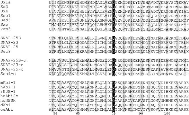
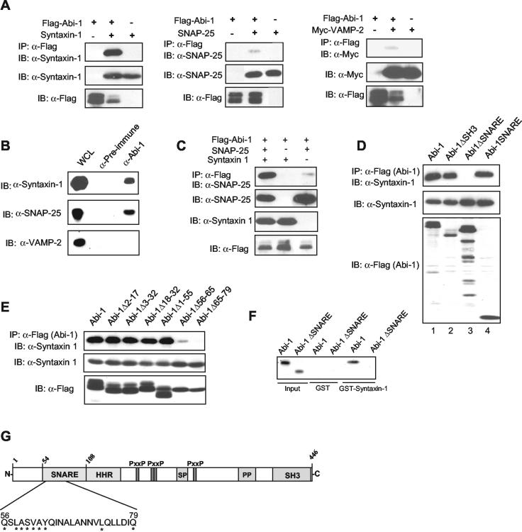
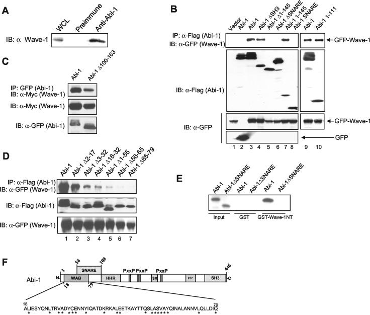
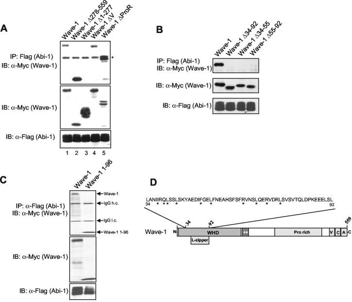
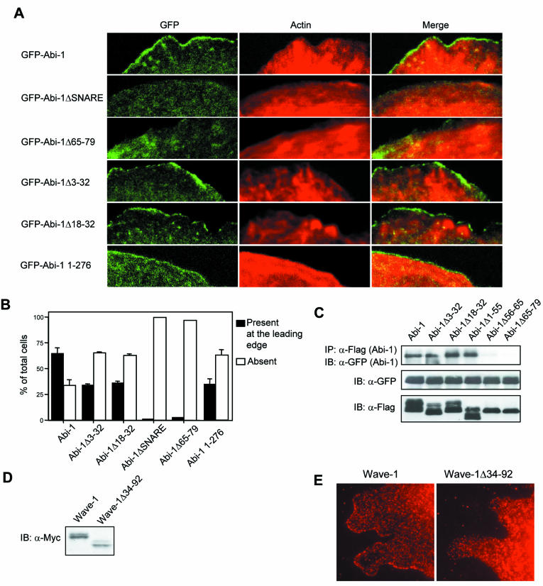
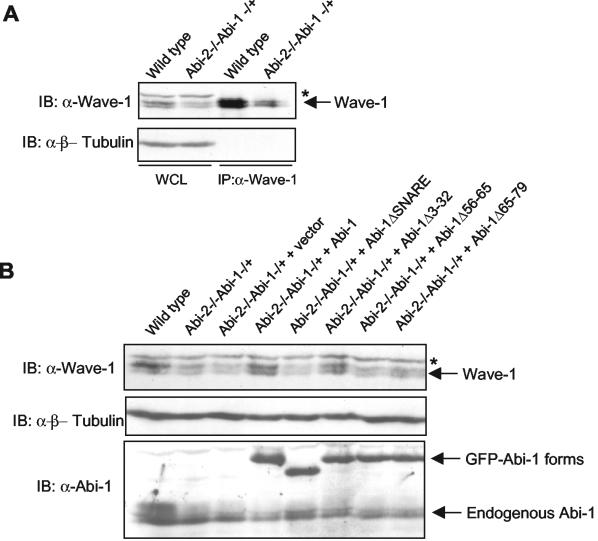
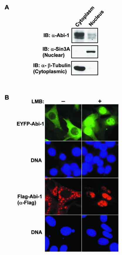
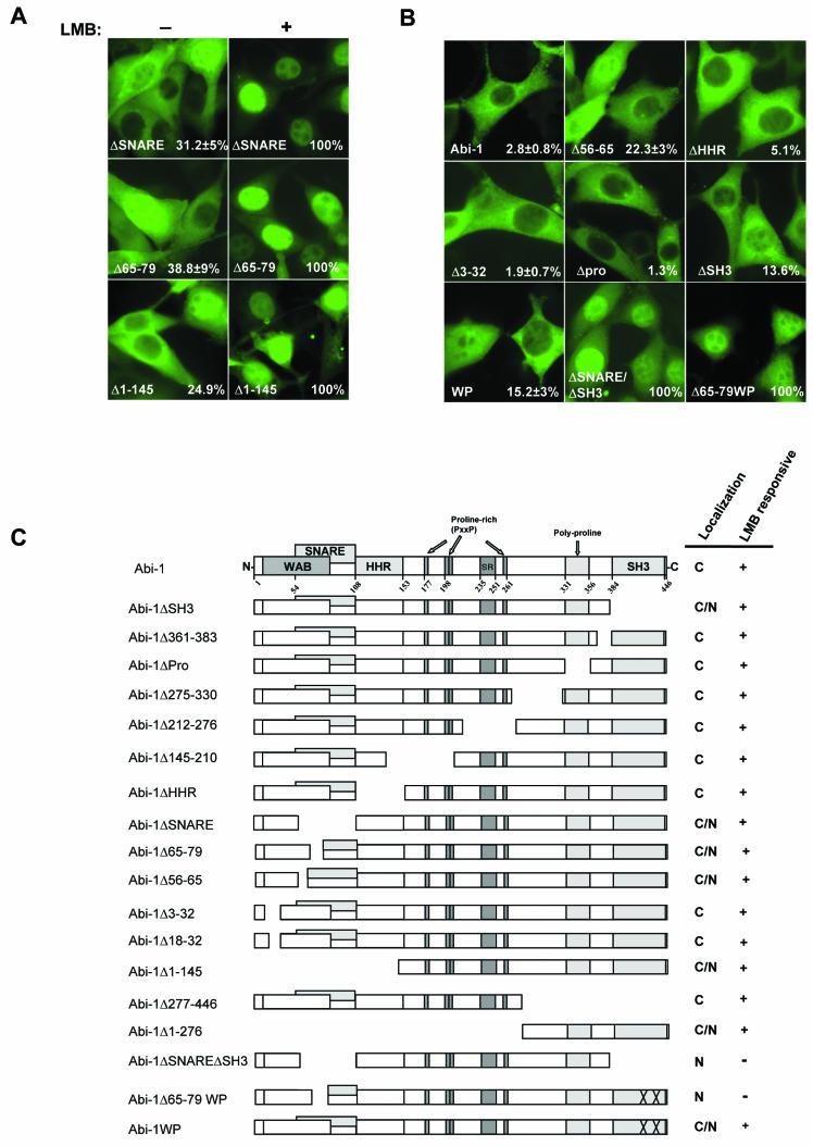
The Abl interactor 1 (Abi-1) protein has been implicated in the regulation of actin dynamics and localizes to the tips of lamellipodia and filopodia. Here, we show that Abi-1 binds the actin nucleator protein Wave-1 through an amino-terminal Wave-binding (WAB) domain and that disruption of the Abi-1-Wave-1 interaction prevents Abi-1 from reaching the tip of the lamellipodium. Abi-1 binds to the Wave homology domain of Wave-1, a region that is required for translocation of Wave-1 to the lamellipodium. Mouse embryo fibroblasts that lack one allele of Abi-1 and are homozygous null for the related Abi-2 protein exhibit decreased Wave-1 protein levels. This phenotype is rescued by Abi-1 proteins that retain Wave-1 binding but not by Abi-1 mutants that cannot bind to Wave-1. Moreover, we uncovered an overlapping SNARE domain in the amino terminus of Abi-1 that interacts with Syntaxin-1, a SNARE family member. Further, we demonstrated that Abi-1 shuttles in and out of the nucleus in a leptomycin B (LMB)-dependent manner and that complete nuclear translocation of Abi-1 in the absence of LMB requires the combined inactivation of the SNARE, WAB, and SH3 domains of Abi-1. Thus, Abi-1 undergoes nucleocytoplasmic shuttling and functions at the leading edge to regulate Wave-1 localization and protein levels.


Figures

FIG. 1.
FIG. 1.
The Abi amino-terminal region exhibits homology to the SNARE motif. Shown is the alignment of Abi family proteins and Q-SNARE family members in the SNARE motif region. Black and gray backgrounds indicate amino acid identity and similarity, respectively. The SNAP-25 family contains two SNARE motifs and the carboxy-terminal motif is indicated by “-c.” Sx, Syntaxin; hu, human; r, rat; m, mouse; d, D. melanogaster; ce, C. elegans. GenBank accession numbers: Sx1a, P32851; Sx3, Q08849; Sx4, Q08850; Sso1, P32867; Sed5, Q01590; Sx5, Q08851; Vam3, Q12241; SNAP-25B, P13795; SNAP-23, U73143; SNAP-25, U81153; Sec9, L34336. NCBI protein accession numbers for the Abi family: mAbi-1, 16225952; hAbi-1, 7839526; rE3B1, 5882255; huAbi2B, 7839524; HuNESH, 14043609; dAbi, 5051971; ceAbi, 17551826. Numbers at the bottom indicate the amino acid positions in hAbi-1.
FIG. 2.
FIG. 2.
Abi-1 binds Syntaxin-1 through the SNARE motif. (A) Abi-1 specifically interacts with Syntaxin-1. Flag-Abi-1 was transfected into 293T cells with Syntaxin-1 (left panel), SNAP-25 (middle panel), or Myc-VAMP-2 (right panel). Abi-1 was immunoprecipitated (IP) with anti-Flag antibodies, and the coprecipitating SNARE proteins were detected by immunoblotting (IB) with the indicated specific antibodies. A DNA ratio of 6:1 was used for Flag-Abi-1 and SNARE proteins. (B) Endogenous Abi-1 interacts with Syntaxin-1. Endogenous Abi-1 was immunoprecipitated from brain extracts with anti-Abi-1 antibody, and the immunopurified complex was immunoblotted with Syntaxin-1, VAMP-2, and SNAP-25 antibodies. Preimmune serum was used as a negative control. (C) Syntaxin-1 bridges the interaction between Abi-1 and SNAP-25. The interaction between SNAP-25 and Abi-1 was detected as described for panel A in the absence or in the presence of Syntaxin-1. (D) The SNARE motif of Abi-1 is necessary and sufficient for interaction with Syntaxin-1 in cells. Flag-tagged full-length Abi-1, Abi-1ΔSH3, Abi-1ΔSNARE, and Abi-1SNARE were transfected with Syntaxin-1-expressing vector into 293T cells, and Abi-1 was immunoprecipitated with anti-Flag antibody. Syntaxin-1 that copurified with Abi-1 is shown in the upper panel, and the levels of Syntaxin-1 and Abi-1 in the total lysates are shown below. (E) Abi-1 amino acids 56 to 79 are critical for interaction with Syntaxin-1. The indicated Flag-Abi-1 deletion mutants were coexpressed with Syntaxin-1, cell lysates were incubated with anti-Flag as described for panel A, and coprecipitating Syntaxin-1 was detected by immunoblotting with anti-Syntaxin-1 antibody. Total lysates were immunoblotted for Syntaxin-1 and Flag-Abi-1 forms. (F) Abi-1 and Syntaxin-1 interact directly. Abi-1 and Abi-1ΔSNARE were synthesized in vitro by using wheat germ extracts and incubated with GST-Syntaxin-1 or GST alone; ∼1% of the total input was loaded in the first two lanes. (G) Schematic representation of the Syntaxin-1-binding region in Abi-1 and the different Abi-1 domains. SR, serine rich; PP, polyproline; P, proline; x, any amino acid. The amino acids required for Syntaxin-1 binding are shown. Asterisks indicate 100% conservation among all Abi-1 family members (human Abi-1, Abi-2, and NESH, as well as mouse Abi-1, D. melanogaster Abi, Anopheles gambiae Abi, and C. elegans Abi).
FIG. 3.
FIG. 3.
Abi-1 interacts with Wave-1 through a conserved amino-terminal WAB domain. (A) Endogenous Abi-1 interacts with Wave-1. Endogenous Abi-1 was immunoprecipitated from brain extracts by using anti-Abi-1 antibody (6987), and the immunopurified complex was immunoblotted (IB) with polyclonal Wave-1 antibody. Preimmune serum was used as a negative control. (B) The region comprising amino acids 1 to 111 of Abi-1 is necessary and sufficient to bind Wave-1. Different Flag-tagged Abi-1 forms and GFP-Wave-1 were coexpressed in 293T cells, and the Flag-Abi-1 forms were immunoprecipitated (IP) by using anti-Flag antibody. In lane 2, GFP was coexpressed with Flag-Abi-1 as a negative control. The coprecipitating GFP-Wave-1 was detected with anti-GFP antibody. Due to low expression levels of Flag-Abi-1SNARE, the ratio of Flag-tagged constructs to GFP-Wave-1 vector was 7:1 in all lanes. (C) EYFP-Abi-1 or EYFP-Abi-1Δ100-163 was cotransfected into 293T cells with Myc-Wave-1, and the Abi-1 forms were immunoprecipitated with anti-GFP antibody. Purified immunocomplexes were immunoblotted with Myc antibody to detect Wave-1 (upper panel). Whole-cell lysates were immunoblotted for Myc and GFP. (D) Abi-1 amino acids 18 to 79 are required for interaction with Wave-1. The indicated Flag-Abi-1 deletion mutants were coexpressed with GFP-Wave-1 and immunoprecipitated with anti-Flag. The coprecipitated GFP-Wave-1 protein was detected with anti-GFP antibodies (upper panel). GFP-Wave-1 protein levels in total cell lysates (lower panel) and the immunoprecipitated Flag-Abi-1 forms (middle panel) were detected with the indicated antibodies. (E) Abi-1 and Wave-1 interact directly. Abi-1 and Abi-1ΔSNARE were synthesized in vitro by using wheat germ extracts and incubated with GST-Wave-1NT or GST alone; ∼5% of the input was loaded in the first two lanes. (F) Schematic representation of the WAB domain in Abi-1. The Abi-1 nomenclature is given in the legend to Fig. 2G.
FIG. 4.
FIG. 4.
Wave-1 interacts with Abi-1 through a region overlapping the putative leucine zipper region. (A) The amino terminus of Wave-1 is necessary for binding to Abi-1. Myc-tagged Wave-1 full-length and Flag-Abi-1 forms were coexpressed in 293T cells, and Flag-Abi-1 forms were immunoprecipitated (IP) with anti-Flag antibodies. Coimmunoprecipitated Wave-1 forms were detected with anti-Myc antibody (upper panel). Expression levels of the Myc-Wave forms are shown in the middle panel, and the levels of Flag-Abi-1 are shown in the lower panel. The asterisk indicates the immunoglobulin G (IgG) heavy-chain band. IB, immunoblot. (B) The region between amino acids 34 to 92 of Wave-1 is necessary to bind Abi-1. Coimmunoprecipitation was performed as described for panel A. The upper panel shows the binding between Flag-Abi-1 and the wild-type and mutant forms of Myc-Wave-1. Wave-1 and Abi-1 levels were visualized by immunoblotting with Myc and Flag antibodies, respectively. (C) Wave-1 amino acids 1 to 96 are sufficient to bind Abi-1. Myc-tagged Wave-1 and Wave-1-1-96 were coexpressed with Flag-Abi-1, and Abi-1 was immunoprecipitated as described for panel A. IgG heavy and light chains are indicated. (D) Schematic representation of the Abi-1-binding region of Wave-1. L-zipper, leucine zipper; ++, basic region; Pro, proline; V, verprolin homology domain; C, cofilin homology domain; A, acidic region. The Wave-1 diagram is not to scale. Asterisks indicate 100% conservation among Wave family members (Wave-1, Wave-2, Wave-3, D. melanogaster Scar, and Dictyostelium discoideum Scar).
FIG. 5.
FIG. 5.
Localization of Abi-1 and Wave-1 to the leading edge of the lamellipodium is dependent on the WAB domain of Abi-1 and the Abi-binding region of Wave-1, respectively. (A) Wild-type MEFs were infected with retroviruses encoding GFP-tagged Abi-1 wild-type and mutant forms, and the Abi-1 proteins were localized by direct observation of the GFP signal (left panels). Actin staining is shown in red (middle panels), and GFP and actin signals are merged in the right panels. Cells were plated for 30 min on fibronectin-coated coverslips, fixed, and visualized by confocal microscopy. (B) Percentages of cells expressing the indicated GFP-Abi-1 forms that exhibit (black bars) or lack (white bars) GFP signal at the leading edge of the lamellipodium. (C) Abi-1 oligomerization depends on the Syntaxin-1-binding region. The indicated Flag-Abi-1 forms and GFP-Abi-1 were coexpressed in 293T cells, and the Flag-Abi-1 proteins were immunoprecipitated (IP) with anti-Flag antibody. Coimmunoprecipitated GFP-Abi-1 was detected with anti-GFP (upper panel). Whole-cell lysates were immunoblotted (IB) with GPF (middle panel) and Flag antibodies (lower panel). (D) Expression of Myc-tagged wild-type Wave-1 and Wave-1Δ34-92 in MEFs. MEFs were infected with retroviruses encoding the indicated Wave-1 proteins, and cell lysates were immunoblotted with anti-Myc antibody. (E) Localization of Wave-1 to the leading edge of the lamellipodium is dependent on the Abi-1-binding region of Wave-1. Wild-type MEFs were infected with a retrovirus encoding Myc-tagged Wave-1 wild type or Wave-1Δ34-92. Cells were plated for 2 h and 15 min on fibronectin-coated coverslips, fixed, and visualized by using anti-Myc antibody.
FIG. 6.
FIG. 6.
Abi-1 and Abi-2 regulate Wave-1 protein levels. (A) Wild-type MEFs or Abi-2−/− Abi-1−/+ MEFs were lysed in KLB, and the lysate was immunoblotted (IB) for Wave-1 and β-tubulin; 1.25 mg of the same lysate was used to immunoprecipitate Wave-1 with anti-Wave-1 monoclonal antibody. (B) Abi-2−/− Abi-1−/+ MEFs were infected with retroviruses expressing the indicated GFP-Abi-1 forms. Cells were lysed as described for panel A and immunoblotted for Wave-1, β-tubulin, and Abi-1. An asterisk indicates a cross-reacting band not present in Wave-1 immunoprecipitates.
FIG. 7.
FIG. 7.
Abi-1 undergoes nucleocytoplasmic shuttling. (A) Endogenous Abi-1 is present in the nucleus. NIH 3T3 cells were lysed, and the lysates were separated into nuclear and cytoplasmic fractions, followed by immunoblotting (IB) with Abi-1 antibody. The purities of nuclear and cytoplasmic fractions were determined by immunoblotting with Sin3A (a nuclear marker) and β-tubulin (a cytoplasmic marker), respectively. (B) Abi-1 accumulates in the nucleus in an LMB-dependent manner. An immunofluorescence signal (green) of EYFP-tagged Abi-1 was observed in transfected NIH 3T3 cells before (−) and after (+) LMB treatment. Flag-tagged Abi-1 in a bicistronic-GFP-expressing vector was expressed following retroviral transduction in NIH 3T3 cells, and Abi-1 localization was determined before and after LMB treatment by immunostaining with Flag monoclonal antibody (red). DAPI staining (DNA; blue) is also shown.
FIG.8.
FIG.8.
The nucleocytoplasmic distribution of Abi-1 is regulated by the Wave-1- and Syntaxin-1-binding regions and the SH3 domain. (A) Cellular localization of the indicated Abi-1 mutants before (−) and after (+) LMB treatment. The number on the bottom right in each panel is the percentage of cells with Abi-1 nuclear staining. (B) Localization of the indicated EYFP-tagged Abi-1 mutants in NIH 3T3 cells in the absence of LMB. The number on the bottom right in each panel is the percentage of cells with Abi-1 nuclear staining. (C) Schematic representation of the Abi-1 mutants employed in this study; their localization is indicated. C, cytoplasmic localization; N, nuclear localization. The Abi-1 nomenclature is given in the legend to Fig. 2G.
FIG. 9.
FIG. 9.
Abi-1 localizes to the leading edge of the lamellipodium and undergoes nucleocytoplasmic shuttling. Distinct pools of Abi-1 bind to Wave-1 or Syntaxin-1 in the cytosol, and localization of Abi-1 to the leading edge of the lamellipodium requires the SNARE and WAB domains in the conserved Abi-1 amino terminus. Disruption of the Abi-1-Wave-1 interaction leads to a reduction of Wave-1 protein levels. Moreover, the binding of Wave-1 to Abi-1 contributes to proper localization of Wave-1 to the leading edge of lamellipodia. Abi-1 undergoes nucleocytoplasmic shuttling and accumulates in the nucleus in response to LMB treatment. Localization of Abi-1 to the nucleus in the absence of LMB requires the simultaneous disruption of the SNARE, WAB, and SH3 domains of Abi-1. These findings suggest that multiple protein-protein interactions are involved in retaining Abi-1 in the cytoplasm. The protein (X) that binds to the Abi-1 SH3 domain and contributes to Abi-1 cytoplasmic retention has not yet been identified. While Wave-1 and Syntaxin-1 bind to the WAB and SNARE domains of Abi-1, respectively, other unknown proteins may also bind to these domains and regulate the nucleocytoplasmic distribution of Abi-1. An unknown factor (Y) is required for the translocation of Abi-1 into the nucleus, and another factor (Z) is required for the nuclear export of Abi-1 through the Crm-1-dependent pathway. YNLS, NLS-containing protein; ZNES, NES-containing protein.

References

    1. Beites, C. L., H. Xie, R. Bowser, and W. S. Trimble. 1999. The septin CDCrel-1 binds syntaxin and inhibits exocytosis. Nat. Neurosci. 5:434-439. - PubMed
    1. Biesova, Z., C. Piccoli, and W. T. Wong. 1997. Isolation and characterization of e3B1, an eps8 binding protein that regulates cell growth. Oncogene 14:233-241. - PubMed
    1. Blagg, S. L., M. Stewart, C. Sambles, and R. H. Insall. 2003. PIR121 regulates pseudopod dynamics and SCAR activity in Dictyostelium. Curr. Biol. 13:1480-1487. - PubMed
    1. Bogerd, H. P., R. A. Fridell, R. E. Benson, J. Hua, and B. R. Cullen. 1996. Protein sequence requirements for function of the human T-cell leukemia virus type 1 Rex nuclear export signal delineated by a novel in vivo randomization-selection assay. Mol. Cell. Biol. 16:4207-4214. - PMC - PubMed
    1. Chapman, E. R., S. An, N. Barton, and R. Jahn. 1994. SNAP-25, a t-SNARE which binds to both syntaxin and synaptobrevin via domains that may form coiled coils. J. Biol. Chem. 269:27427-27432. - PubMed

Publication types