. 2004 Jun;24(11):5028-38.
doi: 10.1128/MCB.24.11.5028-5038.2004.

Cdx1 autoregulation is governed by a novel Cdx1-LEF1 transcription complex

Affiliations

Cdx1 autoregulation is governed by a novel Cdx1-LEF1 transcription complex

Mélanie Béland et al. Mol Cell Biol. 2004 Jun.

Abstract

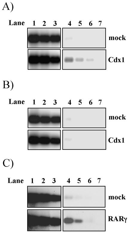
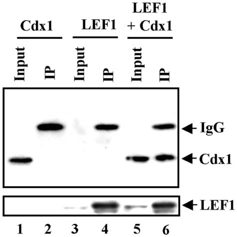
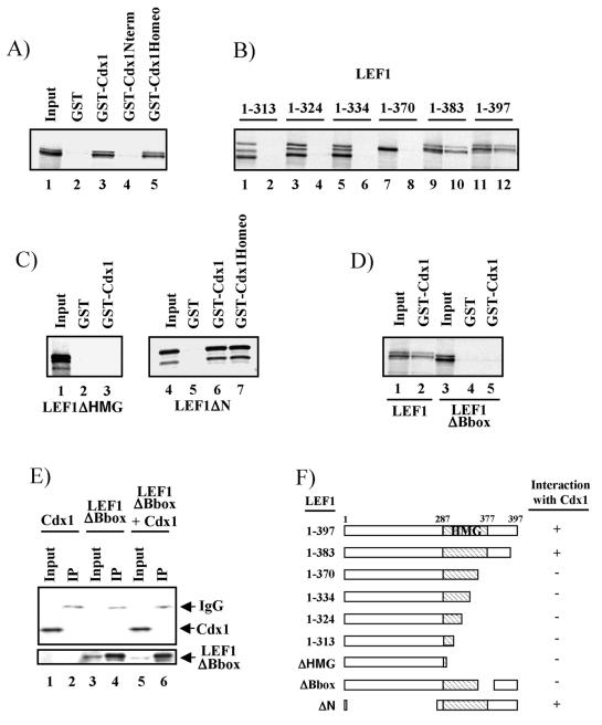
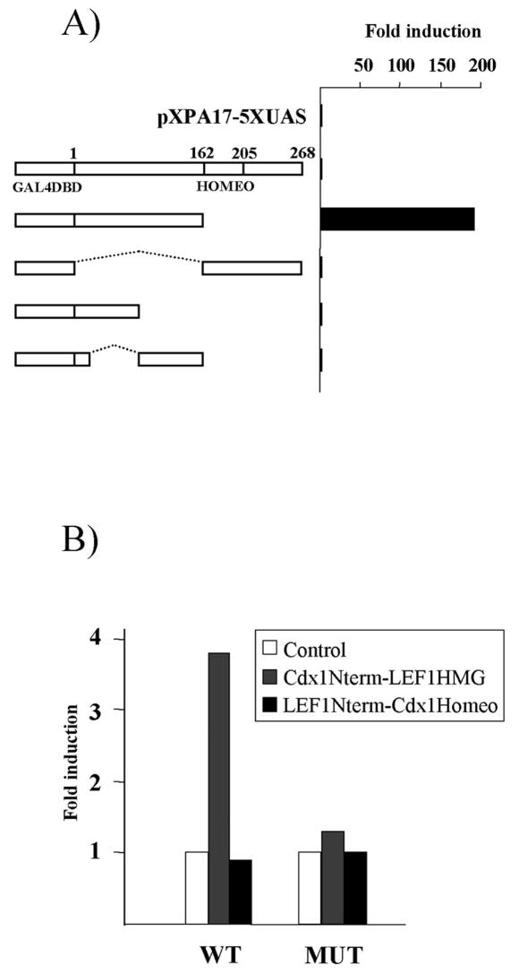
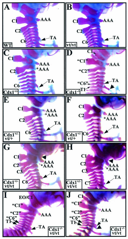
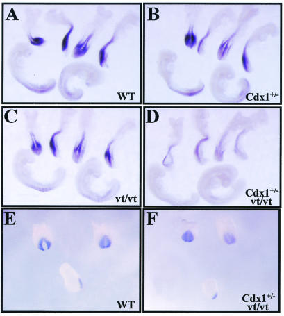
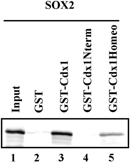
The Cdx1 gene product is essential for normal anterior-posterior vertebral patterning. Expression of Cdx1 is regulated by several pathways implicated in anterior-posterior patterning events, including retinoid and Wnt signaling. We have previously shown that retinoic acid plays a key role in early stages of Cdx1 expression at embryonic day 7.5 (E7.5), while both Wnt3a signaling and an autoregulatory loop, dependent on Cdx1 itself, are involved in later stages of expression (E8.5 to E9.5). This autoregulation is reflected by the ability of Cdx1 to affect expression from proximal Cdx1 promoter sequences in tissue culture. However, this region is devoid of a demonstrable Cdx response element(s). We have now found that Cdx1 and LEF1, a nuclear effector of Wnt signaling, synergize to induce expression from the Cdx1 promoter through previously documented LEF/T-cell factor response elements. We also found a direct physical interaction between the homeodomain of Cdx1 and the B box of LEF1, suggesting a basis for this synergy. Consistent with these observations, analysis of Cdx1 Wnt3a(vt) compound mutants demonstrated that Wnt and Cdx1 converged on Cdx1 expression and vertebral patterning in vivo. Further data suggest that Cdx-high-mobility group box interactions might be involved in a number of additional pathways.


Figures

FIG. 1.
FIG. 1.
Cdx1 is present on its own promoter in vivo. (A and B) Anti-Flag chromatin immunoprecipitation analysis of the endogenous Cdx1 promoter (A) and downstream sequences used as a negative control (B) on mock-transfected (upper panel) and Flag-Cdx1-transfected (lower panel) F9 cells. (C) Identical analysis of the endogenous Cdx1 promoter on mock-transfected (upper panel) and Flag-RARγ (lower panel)-transfected F9 cells. After anti-Flag immunoprecipitation of the sonicated chromatin extracts, DNA was purified and amplified by PCR with oligonucleotides spanning the region of the Cdx1 promoter encompassing the LEF/TCF response elements in panel A, the retinoic acid response element in panel C, or exon 3 in panel B. Products were resolved by agarose gel electrophoresis and assessed by Southern blot analysis with an internal oligonucleotide probe specific to the predicted amplification products. Lanes 1 to 3 (before immunoprecipitation) and 4 to 6 (after immunoprecipitation) represent PCR amplifications of serial dilutions of DNA. Lane 7 is a negative control in which DNA was not included.
FIG. 2.
FIG. 2.
Synergy between Cdx1 and LEF1. (A) P19 cells were transfected with the 2-kb Cdx1 reporter alone or with various amounts of the expression vectors (0.05 to 0.5 μg) as indicated. Cells were harvested 48 h posttransfection, and extracts were assessed for luciferase activity. Data are expressed as induction relative to activity with the reporter vector alone. (B) P19 cells were transfected with a reporter construct harboring Cdx1 binding sites from the Hoxb8 promoter (CDXRE) or with wild-type (pGL3-OT) or mutant (pGL3-OT) LEF/TCF response elements. Transfections were done with the reporter vector alone or with the indicated expression constructs. Luciferase activity was determined as above and expressed as induction relative to activity with the reporter vector alone.
FIG. 3.
FIG. 3.
Cdx1 interacts with LEF1 in vivo. Whole-cell lysates from Cos7 cells transfected with the Cdx1-Flag and/or Lef1-HA expression vectors were immunoprecipitated with anti-HA antibody and analyzed by Western blotting with an anti-Flag antibody (top panel). Expression of LEF1 was assessed by reprobing the blot with an anti-HA antibody (bottom panel). Inputs represent 5% of the total lysate used for immunoprecipitation.
FIG. 4.
FIG. 4.
Cdx1 interacts directly with LEF1. GST-Cdx1 and 35S-labeled LEF1 constructs were generated and assessed for interaction as described in Materials and Methods. (A) Both full-length Cdx1 (GST-Cdx1) and the Cdx1 homeodomain (GST-Cdx1Homeo) interact with LEF1 (lanes 3 and 5), whereas Cdx1 N-terminal sequences do not (lane 4). (B) Deletion of sequences N-terminal to position 383 of LEF1 abolished interaction with Cdx1 (compare lane 10 to lane 8; odd-numbered lanes are 20% input of the various 35S-labeled LEF1 constructs, and even-numbered lanes are from the GST-Cdx1 pulldown). (C) Deletion of the LEF1 HMG box (lane 3) abolishes interaction with Cdx1, whereas loss of N-terminal LEF1 residues has no effect on interaction with either full-length Cdx1 (lane 6) or the homeodomain sequences of Cdx1 (lane 7). (D and E) Deletion of the LEF1 B box abolishes interaction with Cdx1 in vitro (D, compare lane 5 to lane 2) and in vivo (E, lane 6). (F) Schematic representation of LEF1-Cdx1 interactions. Coimmunoprecipitations (panel E) were performed as in Fig. 2 except the ΔBbox mutant was used instead of full-length LEF1.
FIG. 5.
FIG. 5.
Transcriptional activation domain of Cdx1 activates the Cdx1 promoter. (A) The N-terminal domain of Cdx1 contains a transactivation function. P19 cells were transfected with a reporter vector containing GAL4 binding elements (pXPA17-5XUAS), alone or with the indicated GAL4 DNA binding domain-Cdx1 fusion. Luciferase activity was measured 48 h posttransfection and expressed as induction relative to activity with the reporter vector alone. (B) The N-terminal domain of Cdx1 can activate transcription from the Cdx1 promoter when fused to the LEF1 HMG box. P19 cells were transfected with the 180-bp proximal Cdx1 reporter construct (WT) or an identical construct lacking the LEF/TCF response elements (MUT). Transfections were performed either with the reporter vector alone or with the indicated fusion construct. Luciferase activity and results were determined as above.
FIG. 6.
FIG. 6.
Skeletal analysis of Cdx1 Wnt3avt compound mutants. The cervical region of whole-mount skeletal preparations from (A) wild-type, (B) Wnt3avt/vt, (C) Cdx1+/−, (D) Cdx1−/−, (E and F) Cdx1+/− Wnt3avt/+, (G and H) Cdx1+/− Wnt3avt/vt and (I and J) Cdx1−/− Wnt3avt/vt offspring. Note the normal cervical region for both wild-type (A) and Wnt3avt/vt (B) mice. (C, E, and F) Cdx1+/− and Cdx1+/− Wnt3avt/+ mice exhibit similar phenotypes, notably an ectopic anterior arch of the atlas (*AAA), a broader C2 neural arch, and a C1-C2 fusion (F). (D, I, and J). Cdx1 null and Cdx1−/− Wnt3avt/vt mice exhibit identical vertebral defects, notably malformation and malposition of C1 and anterior transformation of C2, C3, and C7, denoted “C1,” “C2,” and “C6,” respectively. The arrowhead in panel J indicates a partial rib associated with presumptive T1 which does not reach the sternum. (G and H) Cdx1+/− Wnt3avt/vt offspring exhibit an exacerbated phenotype compared to either single mutant (compare panels G and H to panels B and C). In particular, note the fusion of the anterior arch of the atlas with the basioccipital bone (arrowhead) and the presence of an ectopic anterior arch of the atlas. Note also the malformations and fusions of neural arches C1 to C3 and the presence of TA on C7 instead of C6 (H). Abbreviations: AAA, anterior arch of the atlas; *AAA, ectopic anterior arch of the atlas; TA, tuberculum anterior; EO, exoccipital; C, cervical vertebrae; T, thoracic vertebrae. Quotation marks indicate presumptive anterior transformations.
FIG. 7.
FIG. 7.
Stage-specific attenuation of Cdx1 expression in Cdx1+/− Wnt3avt/vt compound mutants. Cdx1 expression was assessed by whole-mount in situ hybridization in E8.5 (A to D) or E7.5 (E and F) embryos. (A and E) Wild-type control. (B) Cdx1+/−. (C) Wnt3avt/vt. (D and F) Cdx1+/− Wnt3avt/vt. Note the nearly complete loss of Cdx1 expression at E8.5 in Cdx1+/− Wnt3avt/vt embryos relative to that in the cognate mutant backgrounds, while expression is unaffected at E7.5.
FIG. 8.
FIG. 8.
Cdx1 homeodomain interacts with Sox2 in vitro. 35S-labeled Sox2 was incubated with GST alone or GST-Cdx1 (full length, N terminus, or homeodomain only). Sox2 specifically interacted with both full-length Cdx1 (lane 3) and the Cdx1 homeodomain (lane 5).

References

    1. Allan, D., M. Houle, N. Bouchard, B. I. Meyer, P. Gruss, and D. Lohnes. 2001. RAR gamma and Cdx1 interactions in vertebral patterning. Dev. Biol. 240:46-60. - PubMed
    1. Beck, F., T. Erler, A. Russell, and R. James. 1995. Expression of Cdx-2 in the mouse embryo and placenta: possible role in patterning of the extra-embryonic membranes. Dev. Dyn. 204:219-227. - PubMed
    1. Bel-Vialar, S., N. Itasaki, and R. Krumlauf. 2002. Initiating Hox gene expression: in the early chick neural tube differential sensitivity to FGF and RA signaling subdivides the HoxB genes in two distinct groups. Development 129:5103-5115. - PubMed
    1. Boras, K., and P. A. Hamel. 2002. Alx4 binding to LEF-1 regulates N-CAM promoter activity. J. Biol. Chem. 277:1120-1127. - PubMed
    1. Burke, A. C., C. E. Nelson, B. A. Morgan, and C. Tabin. 1995. Hox genes and the evolution of vertebrate axial morphology. Development 121:333-346. - PubMed

Publication types

MeSH terms

LinkOut - more resources