. 2004 Jun;5(6):579-83.
doi: 10.1038/sj.embor.7400149. Epub 2004 May 14.

Following single antibody binding to purple membranes in real time

Affiliations

Following single antibody binding to purple membranes in real time

Ferry Kienberger et al. EMBO Rep. 2004 Jun.

Abstract

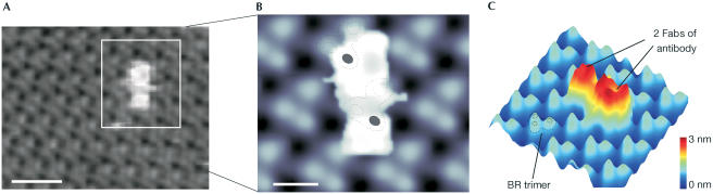
Antibody binding to surface antigens in membranes is the primary event in the specific immune defence of vertebrates. Here we used force microscopy to study the dynamics of antibody recognition of mutant purple membranes from Halobacterium salinarum containing a genetically appended anti-Sendai recognition epitope. Ligation of individual anti-Sendai antibodies to their antigenic epitopes was observed over time. Their increase in number within a small selected area revealed an apparent kinetic on-rate. The membrane-bound antibodies showed many different conformations that ranged from globular to V- and Y-like shapes. The maximum distance of two Fab fragments of the same antibody was observed to be approximately 18 nm, indicating an overall strong intrinsic flexibility of the antibody hinge region. Fab fragments of bound anti-Sendai antibodies were allocated to antigenic sites of the purple membrane, allowing the identification and localization of individual recognition epitopes on the surface of purple membranes.


Figures

Figure 1
Figure 1
Kinetics of antisendai antibody binding to Sendai PM. (A) Schematic representation of a Sendai BR molecule. In contrast to a wild-type BR molecule, Sendai BR has a 13-amino-acid-long Sendai epitope genetically fused in the cytoplasmic E–F loop. (B) Specificity of the anti-Sendai antibody–antigen interaction. The specificity of the antibody–antigen interaction is shown on AFM topography images. The left image shows anti-Sendai antibodies (visible as bright dots) covering Sendai PM after 20 min of incubation. No anti-Sendai antibodies were detected on wild-type PM even after 2 h incubation time (right image). Scan size was 500 nm on both images. Scale bar, 150 nm. (C) Successive AFM topography images of Sendai PM at (0 min) and after (48 and 72 min) antibody incubation. Upon binding to PM, they appear as bright structures (see arrows) 2–3 nm in height (see height false colour bar, 0–8 nm). Scan size was 300 nm on all three images. Scale bar, 100 nm. (D) Diagram depicting the increase in the number of bound antibodies (nAbBR) in the image frame of (C) with incubation time. An initial lag phase (from 0 to 40 min) is followed by a linear increase in the number of membrane-bound antibodies (from 48 to 88 min).
Figure 2
Figure 2
Substructures of antibodies specifically bound to Sendai PM. (A) Large membrane patches of Sendai PM covered with antibodies. The white box indicates the region that was enlarged for (B). Antibodies appear as small dots 10–20 nm in diameter and 2–3 nm in height. Scan size was 1 μm. Scale bar, 200 nm. (B) Structural details of the membrane-attached antibodies obtained in smaller scan areas. Several different arrangements of Fab fragments can be easily discerned. Scan size was 150 nm. Scale bar, 40 nm. (C) Classes of antibody conformations. Antibody shapes were divided into three major classes, corresponding to their Fab and Fc arrangement: (i) globular, (ii) and (iii) Vshaped, and (iv) Y-shaped. The lower-right sketch shows the three-dimensional morphology and size of an antibody (Silverton et al, 1977). Image size was 35 nm in all four images. Scale bar, 20 nm.
Figure 3
Figure 3
Localization of antibody-binding epitopes on Sendai PM. (A) Topographical image of the cytoplasmic side of Sendai PM to which an antibody is bound. The hexagonal lattice structure of the membrane can be discerned over the whole scan size in this Fourier-filtered image. The white box indicates the region for subsequent analysis. Image size was 40 nm. Scale bar,10 nm. (B) The hexagonal lattice structure of BR trimers and the arrangement of the two Fab fragments of the antibody are clearly visible at higher magnification. The dotted contour lines depict BR trimers covered by the antibody. The BR molecules that are engaged by the two Fab fragments of the antibody are marked in black. Image size was 20 nm. Scale bar, 4 nm. (C) Three-dimensional presentation of (B) showing the heights of the structures. The BR trimers (indicated with dotted lines) protrude 0.8 nm out of the lipid bilayer, whereas the two Fab fragments (see labelling) of the antibody are 2–3 nm in height.

References

    1. Butt HJ, Jaschke M (1995) Thermal noise in atomic force microscopy. Nanotechnology 6: 1–7
    1. Cheung CL, Hafner JH, Lieber CM (2000) Carbon nanotube atomic force microscopy tips: direct growth by chemical vapor deposition and application to high-resolution imaging. Proc Natl Acad Sci USA 97: 3809–3813 - PMC - PubMed
    1. Einberger H, Mertz R, Hofschneider PH, Neubert WJ (1990) Purification, renaturation, and reconstituted protein kinase activity of the Sendai virus large (L) protein: L protein phosphorylates the NP and P proteins in vitro. J Virol 64: 4274–4280 - PMC - PubMed
    1. Gropp F, Gropp R, Betlach MC (1995) Effects of upstream deletions on light-and oxygen-regulated bacterio-opsin gene expression in Halobacterium halobium. Mol Microbiol 16: 357–364 - PubMed
    1. Han W, Mou J, Sheng J, Yang J, Shao Z (1995) Cryo atomic force microscopy: a new approach for biological imaging at high resolution. Biochemistry 34: 8215–8220 - PubMed

Publication types

MeSH terms