

FMRI of brain activation in a genetic rat model of absence seizures
- PMID: 15144421
- PMCID: PMC2949946
- DOI: 10.1111/j.0013-9580.2004.39303.x
FMRI of brain activation in a genetic rat model of absence seizures
Abstract
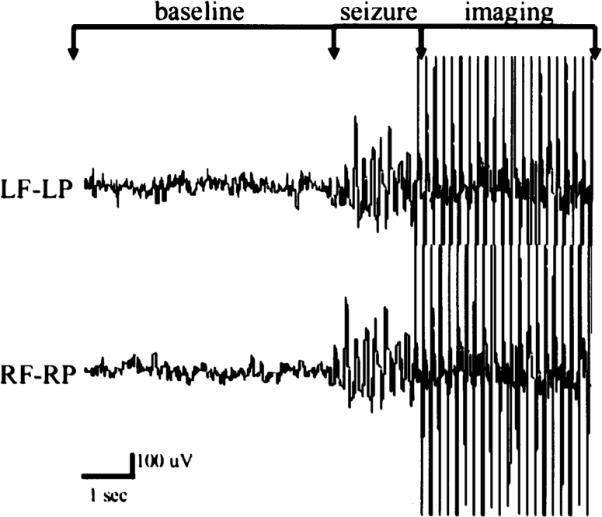
Purpose: EEG-triggered functional magnetic resonance imaging (fMRI) was used to identify areas of brain activation during spontaneous spike-and-wave discharges (SWDs) in an epileptic rat strain under awake conditions.
Methods: Spontaneous absence seizures from 10 WAG/Rij rats were imaged by using T2*-weighted echo planar imaging at 4.7 Tesla. fMRI of the blood-oxygenation-level-dependent (BOLD) signal was triggered based on EEG recordings during imaging. Images obtained during spontaneous SWDs were compared with baseline images.
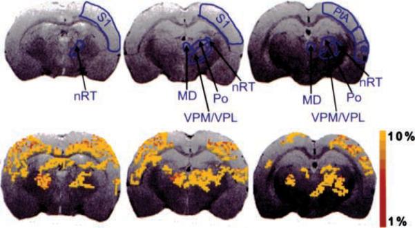
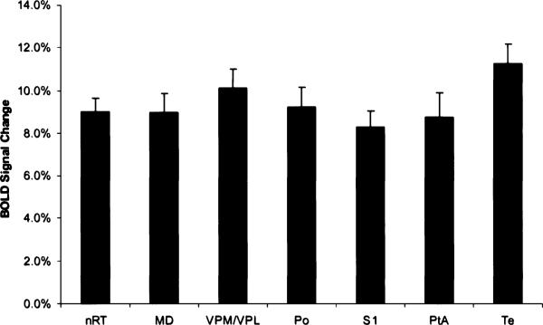
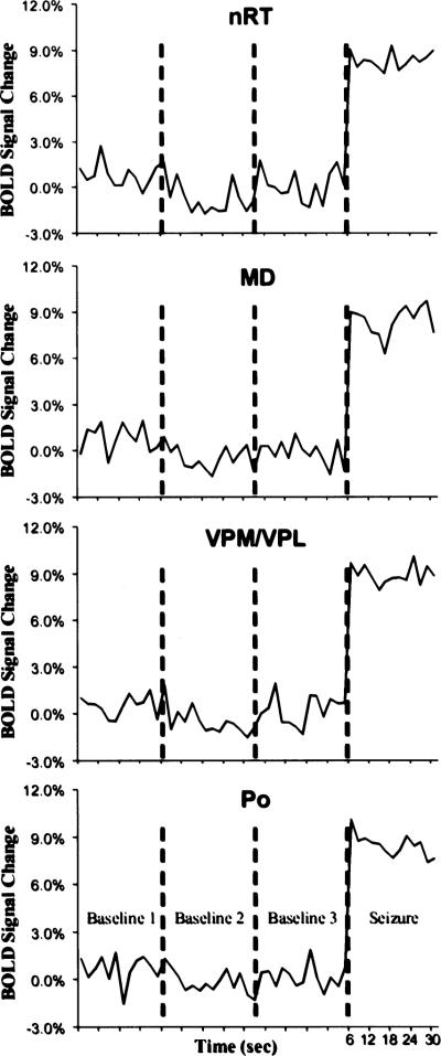
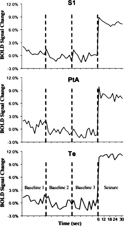
Results: Significant positive BOLD signal changes were apparent in several areas of the cortex and several important nuclei of the thalamus. In addition, no negative BOLD signal was found in any brain area.
Conclusions: We have shown that EEG-triggered BOLD fMRI can be used to detect cortical and thalamic activation related to the spontaneous SWDs that characterize absence seizures in awake WAG/Rij rats. These results draw an anatomic correlation between areas in which increased BOLD signal is found and those in which SWDs have been recorded. In addition, no negative BOLD signal was found to be associated with these spontaneous SWDs. We also demonstrated the technical feasibility of using EEG-triggered fMRI in a genetic rat model of absence seizure.
Figures
Similar articles
-
Increased resting functional connectivity in spike-wave epilepsy in WAG/Rij rats.Epilepsia. 2013 Jul;54(7):1214-22. doi: 10.1111/epi.12227. Epilepsia. 2013. PMID: 23815571 Free PMC article.
-
Corticothalamic modulation during absence seizures in rats: a functional MRI assessment.Epilepsia. 2003 Sep;44(9):1133-40. doi: 10.1046/j.1528-1157.2003.61002.x. Epilepsia. 2003. PMID: 12919383 Free PMC article.
-
fMRI of generalized absence status epilepticus in conscious marmoset monkeys reveals corticothalamic activation.Epilepsia. 2004 Oct;45(10):1240-7. doi: 10.1111/j.0013-9580.2004.21504.x. Epilepsia. 2004. PMID: 15461678
-
Spike-wave discharges in adult Sprague-Dawley rats and their implications for animal models of temporal lobe epilepsy.Epilepsy Behav. 2014 Mar;32:121-31. doi: 10.1016/j.yebeh.2014.01.004. Epub 2014 Feb 15. Epilepsy Behav. 2014. PMID: 24534480 Free PMC article. Review.
-
Pathophysiological mechanisms of genetic absence epilepsy in the rat.Prog Neurobiol. 1998 May;55(1):27-57. doi: 10.1016/s0301-0082(97)00091-9. Prog Neurobiol. 1998. PMID: 9602499 Review.
Cited by
-
Simultaneous fMRI and local field potential measurements during epileptic seizures in medetomidine-sedated rats using raser pulse sequence.Magn Reson Med. 2010 Oct;64(4):1191-9. doi: 10.1002/mrm.22508. Magn Reson Med. 2010. PMID: 20725933 Free PMC article.
-
Tissue hypoxia correlates with intensity of interictal spikes.J Cereb Blood Flow Metab. 2011 Jun;31(6):1394-402. doi: 10.1038/jcbfm.2011.16. Epub 2011 Feb 23. J Cereb Blood Flow Metab. 2011. PMID: 21343943 Free PMC article.
-
Connecting the dots between cell populations, whole-brain activity, and behavior.Neurophotonics. 2022 Jul;9(3):032208. doi: 10.1117/1.NPh.9.3.032208. Epub 2022 Mar 26. Neurophotonics. 2022. PMID: 35350137 Free PMC article.
-
Rhythmic 3-4Hz discharge is insufficient to produce cortical BOLD fMRI decreases in generalized seizures.Neuroimage. 2015 Apr 1;109:368-77. doi: 10.1016/j.neuroimage.2014.12.066. Epub 2015 Jan 3. Neuroimage. 2015. PMID: 25562830 Free PMC article.
-
Effects of Hemodynamic Response Function Selection on Rat fMRI Statistical Analyses.Front Neurosci. 2019 Apr 30;13:400. doi: 10.3389/fnins.2019.00400. eCollection 2019. Front Neurosci. 2019. PMID: 31114471 Free PMC article.
References
-
- Festing MFW. Inbred strains in biomedical research. MacMillan Press; London: 1979. pp. 267–96.
-
- van Luijtelaar ELMJ, Coenen AML. Two types of electrocortical paroxysms in an inbred strain of rats. Neurosci Lett. 1986;70:393–7. - PubMed
-
- Coenen AML, Drinkenburg WHIM, Inoue M, et al. Genetic models of absence epilepsy, with emphasis on the WAG/Rij strain of rats. Epilepsy Res. 1992;12:75–86. - PubMed
-
- Coenen AML, van Luijtelaar ELJM. The WAG/Rij rat model for absence epilepsy: age and sex factors. Epilepsy Res. 1987;1:297–301. - PubMed
Publication types
MeSH terms
Substances
Grants and funding
LinkOut - more resources
Full Text Sources
Medical
