. 2004 Jul 15;558(Pt 2):577-96.
doi: 10.1113/jphysiol.2004.063586. Epub 2004 May 14.

Mechanosensory S-neurons rather than AH-neurons appear to generate a rhythmic motor pattern in guinea-pig distal colon

Affiliations

Mechanosensory S-neurons rather than AH-neurons appear to generate a rhythmic motor pattern in guinea-pig distal colon

Nick J Spencer et al. J Physiol. .

Abstract

Simultaneous intracellular recordings were made from myenteric neurons and circular muscle (CM) cells in isolated, stretched segments of guinea-pig distal colon. We have shown previously that maintained stretch generates a repetitive and coordinated discharge of ascending excitatory and descending inhibitory neuronal reflex pathways in the distal colon. In the presence of nifedipine (1-2 microm) to paralyse the muscle, simultaneous recordings were made from 25 pairs of AH (after-hyperpolarization)-neurons and CM cells separated by 100-500 microm. In all 25 AH-neurons, proximal process potentials (PPPs) were never recorded, even though at the same time, all recordings from neighbouring CM cells showed an ongoing discharge of inhibitory junction potentials (IJPs) anally, or excitatory junction potentials (EJPs) orally. In fact, 24 of 25 AH-neurons were totally silent, while in one AH-cell, some spontaneous fast excitatory postsynaptic potentials (FEPSPs) were recorded. All 10 electrically silent AH-cells that were injected with neurobiotin were found to be multipolar Dogiel type II neurons. In contrast, when recordings were made from myenteric S-neurons, two distinct electrical patterns of electrical activity were recorded. Recordings from 25 of 48 S-neurons showed spontaneous FEPSPs, the majority of which (22 of 25) showed periods when discrete clusters of FEPSPs (mean duration 88 ms) could be temporally correlated with the onset of EJPs or anal IJPs in the CM. Nine S-neurons were electrically quiescent. The second distinct electrical pattern in 14 S-neurons consisted of bursts, or prolonged trains of action potentials, which could be reduced to proximal process potentials (PPPs) in six of these 14 neurons during membrane hyperpolarization. Unlike FEPSPs, PPPs were resistant to a low Ca(2+)-high Mg(2+) solution and did not change in amplitude during hyperpolarizing pulses. Mechanosensory S-neurons were found to be uniaxonal or pseudounipolar filamentous neurons, with morphologies consistent with interneurons. No slow EPSPs were ever recorded from AH- or S-type neurons when IJPs or EJPs occurred in the CM. In summary, we have identified a population of mechanosensory S-neurons in the myenteric plexus of the distal colon which appear to be largely stretch sensitive, rather than muscle-tension sensitive, since they generate ongoing trains of action potentials in the presence of nifedipine. No evidence was found to suggest that in paralysed preparations, the repetitive firing in ascending excitatory or descending inhibitory nerve pathways was initiated by myenteric AH-neurons, or slow synaptic transmission.


Figures

Figure 1
Figure 1. Inactivity in a myenteric AH neuron during ascending excitation
A, diagrammatic representation of the preparation used for simultaneous recording from myenteric neurons and CM cells in preparations where the CM was intact, but the LM removed (CM-MP preparation; see Methods). B shows a typical simultaneous recording from a myenteric AH-neuron and CM cell located < 500 μm apart and within 1 mm of the oral cut end of colon. An ongoing discharge of EJPs occurs in the CM at the oral end of the colon. At the same time as EJPs occurred, this AH-neuron in a neighbouring ganglia was electrically quiescent, until depolarizing current was injected into the neuron on two occasions to evoke action potentials. Following the evoked action potentials a prolonged membrane hyperpolarization ensued. Evoked action potentials in this neuron did not cause any detectable membrane potential change in the CM cell. C shows that the AH-neuron recorded in B was a multipolar Dogiel type II neuron. D, an expanded portion of the recording shown in B, showing that the evoked action potentials in this neuron did not change CM membrane potential. The calibration bar in C represents 30 μm. The resting membrane potentials of the CM cell and the AH-neuron were −45 and −61 mV.
Figure 2
Figure 2. Morphology and projections of an electrically silent myenteric AH-neuron recorded from the anal end of distal colon in a CM-MP preparation
A, diagrammatic representation of the relative locations of the two recording electrodes at the anal end of colon. The LM was removed while the CM remained intact. B shows a simultaneous recording from an AH-neuron and a closely apposed CM cell (500 μm apart). An ongoing discharge of large amplitude IJPs occurred in the CM cell, even though at the same time the AH-neuron was silent. Depolarizing current injection evoked two action potentials in the soma, followed by a prolonged membrane after-hyperpolarization. C shows an expanded portion of the evoked action potential shown in B. D, the neuron in A was found to be a multipolar Dogiel type II neuron (see arrow) with a long process that appeared to enter the CM layer. E, the cell body of the AH-neuron from which the recording was made (see arrow in D). F shows an enlarged image of the nerve endings in the CM layer. The cell body was measured 40 μm (major axis) and 23 μm (minor axis). The calibration bars in DF represent 25 μm. The resting membrane potentials of the CM cell and the AH-neuron were −49 and −62 mV.
Figure 3
Figure 3. Electrical activity recorded from a mechanosensory S-neuron in the myenteric plexus, when pinned under maintained circumferential stretch
A, spontaneous action potentials and proximal process potentials occurred in this neuron when recorded in the presence of nifedipine, and were unaffected by the application of a low Ca2+ –high Mg2+ solution to block all synaptic transmission (see hatched bar). B shows an expanded portion of the recording in the low Ca2+ –high Mg2+ solution. Note, the similarity in amplitude of process potentials recorded in the low Ca2+ solution. Some process potentials were sufficient to trigger a full somatic action potential. C, some spontaneous FEPSPs were recorded in this S-neuron prior to the application of the low Ca2+ –high Mg2+ solution. These are identified by their irregular amplitudes (see arrow). D, this neuron fired tonically in response to depolarizing current. E, in the presence of the low Ca2+ –high Mg2+ solution, removal of hyperpolarizing holding current increased the discharge of process potentials, suggesting that some mechanosensory ion channels may exist close to, or even in the cell soma itself. F, this neuron was an orally projecting uniaxonal filamentous neuron. The calibration bar represents 20 μm. The resting potential of this neuron was −53 mV.
Figure 4
Figure 4. Simultaneous recording from a mechanosensory S-neuron and a circular muscle cell
A, ongoing IJPs occur in the CM cell at the same time as action potentials discharge spontaneously in this S-neuron. No correlation was observed between spontaneous action potentials in this S-neuron and the IJPs in the CM. When a conditioning hyperpolarization of ∼50 mV was imposed on the cell soma, action potentials were converted into proximal process potentials. When the hyperpolarizing current was withdrawn, process potentials were converted back to full somatic action potentials. B, in response to depolarizing current, this was a tonic firing cell, but the train of action potentials did not cause any change in membrane potential in the neighbouring CM. C, in addition to process potentials and spontaneous action potentials, this neuron also received prominent FEPSPs, distinguishable by their irregular amplitudes. D shows the filamentous processes of this Dogiel type I neuron. This neuron appeared to be uniaxonal and projected anally for at least one row of ganglia, but was not filled sufficiently to trace its ending. The calibration bar represents 20 μm. The resting membrane potential of this neuron was −50 mV.
Figure 5
Figure 5. Intrinsically active ascending interneuron in a stretched segment of distal colon
A, ongoing action potentials and membrane noise recorded from a stretched preparation, while in the presence of nifedipine. Note the membrane noise during the interspike intervals. B, ongoing action potentials and noise are abolished by membrane hyperpolarization. This suggests that the action potentials arise close to or within the cell soma. C, morphology of this ascending interneuron, showing fine varicose synaptic outputs in the first and second rows of ganglia (see boxes 1 and 2, shown on enlarged scale in E and F). This neuron had a long circumferentially projecting dendrite that arose from the soma. D, enlarged image of the neuronal soma and processes. E and F show the synaptic outputs of this ascending interneuron, expanded from boxes 1 and 2 in C. G and H show a short process that appears to enter the CM layer. G, when the soma is in focus, the end of this process is out of focus, suggesting it is not in the plane of the soma. H shows this process in focus (see arrow) and entering the CM layer, while the soma is now out of focus compared with G. The long axon of this neuron eventually left a ganglion (most oral ganglion shown in the top panel in C) and entered and dived down through the CM ending in an expansion bulb on the submucosal surface of the CM (not shown), suggesting that it may terminate in the submucous plexus or the mucosa. Calibration bars represent 100 μm in C, 35 μm in D, 13 μm in E and F, and 17 μm in G and H.
Figure 6
Figure 6. Electrical and morphological characteristics of a mechanosensory descending interneuron
A, morphology of a pseudounipolar descending neuron that gave rise to two axons (more oral arrow). The thin axon appeared to provide synaptic outputs in the second row of ganglia (more anal arrow). The thicker darker axon appeared to leave a ganglia and enter the neighbouring CM (see *). B shows the cell body of this neuron with filamentous dendritic processes, one of which runs parallel to the CM fibres. C, this neuron had two bifurcating axons close to the axon hillock; see arrow. D, spontaneous proximal process potentials and action potentials occurred in this neuron under stretch and in the presence of nifedipine. When the membrane was artificially hyperpolarized by ∼50 mV the amplitude and interval between process potentials did not change, suggesting that these events were electrotonically invading the soma from a site distant to the cell body. E shows removal of hyperpolarizing holding current, where process potentials do not change characteristics. F, when simultaneous recordings were made from a CM cell and an S-neuron, single pulse transmural stimulation evoked FEPSPs (see arrow) and a single action potential, followed by a fast IJP in the CM. G, in addition to proximal process potentials, this neuron showed ongoing spontaneous fast EPSPs. These were identified because their amplitudes were highly variable. H, this neuron fired action potentials tonically in response to depolarizing current injection. The resting membrane potential of this neuron was −48 mV.
Figure 7
Figure 7. Properties of myenteric S-neurons in stretched preparations of distal colon while in the presence of nifedipine
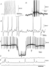
A, a tonic S-neuron firing in response to depolarizing current. Ai, while in the presence of a low Ca2+ –high Mg2+ solution, the neuron shown in A responded with a burst of action potentials in response to a ganglionic compression stimulus to the CM. B, in a different animal, an intrinsically active S-neuron showed an ongoing discharge of membrane noise represented by the unstable membrane potential. C, effects of membrane hyperpolarization on an S-neuron that had spontaneous action potentials and fast EPSPs. When the membrane potential was artificially hyperpolarized, the action potentials ceased, and only fast EPSPs were recorded. Fast EPSPs were detected because they increased in amplitude and had irregular amplitudes. Compare panels b, c and d, shown on an expanded scale in DF. Recordings shown in AC are all from different animals.
Figure 8
Figure 8. Morphology and synaptic inputs to a myenteric S-neuron involved in repetitively discharging ascending excitatory nerve pathways
A, simultaneous recording from a myenteric S-neuron and CM at the oral end of a CM-MP preparation. Prior to the onset of each EJP in the CM, a brief burst of fast EPSPs occurred in this neuron. Note that during the IJPs that occurred in the CM, this S-neuron did not receive fast EPSPs, suggesting that it may have been an excitatory motor neuron. B, this neuron was injected with neurobiotin and found to be a uniaxonal Dogiel type I neuron with short lamellar dendrites. C, an expanded image of the cell body of the neuron recorded from in A. D shows another simultaneous recording from an S-neuron and CM cell in a different animal. Note that prior to each EJP a brief burst of fast EPSPs occurred in the S-cell. E, the recording period represented by the bar in D is shown on an expanded time scale. Fast EPSPs and the single EJP are shown. Note that the discrete burst of fast EPSPs immediately precedes the EJP by approximately 150 ms. The calibration bar represents 40 μm in B and 15 μm in C. The resting membrane potentials of the CM cell and the S-neuron were −36 and −45 mV.
Figure 9
Figure 9. Firing patterns of a myenteric S-neuron during repetitively discharging ascending excitatory nerve pathways
A, simultaneous recording from an S-neuron and CM cell at the oral end of a CM-MP preparation. Note that the duration of the fast EPSPs and action potentials that precede each EJP are similar, but the amplitudes of each EJP in the CM are dissimilar. B, expanded trace from the period represented by the black bar in A. C, this neuron fired a brief burst of action potentials in response to depolarizing current. The resting membrane potential of this neuron was −47 mV.
Figure 10
Figure 10. Characteristics of fast synaptic potentials in a myenteric S-neuron that underlies EJP generation in circular muscle
A and B show a simultaneous recordings from the same S-neuron and CM cell as in Fig. 9 at the oral end of a CM-MP preparation. Note the EJPs vary widely in amplitude in A and B, yet the duration of the fast EPSP burst in the S-neuron is similar. Arrows a and b (see A) indicate how the duration of FEPSPs and the latency of EJP onset were measured. C, recordings from A and B are superimposed to show the similarity in duration of fast EPSP bursts, despite large differences in EJP amplitude. D, summarized data from 26 different S-neurons showing that there was no temporal correlation between the duration of the fast synaptic inputs in an individual S-neuron compared with the amplitude of the EJP in the CM (R2 = 0.01). E, similarly, there was no correlation between the latency of a fast EPSP burst in an S-neuron (see arrow b in A) when compared to the amplitude of an EJP in the CM (R2 = 0.005). The resting membrane potential of this neuron was −47 mV.

Similar articles

Cited by

References

    1. Bayliss WM, Starling EH. The movements and innervation of the small intestine. J Physiol. 1899;24:99–143. - PMC - PubMed
    1. Bayliss WM, Starling EH. The movements and innervation of the large intestine. J Physiol. 1900;26:107–118. - PMC - PubMed
    1. Bertrand PP, Bornstein JC. ATP as a putative sensory mediator: activation of intrinsic sensory neurons of the myenteric plexus via P2X receptors. J Neurosci. 2002;22:4767–4775. - PMC - PubMed
    1. Bornstein JC, Furness JB, Smith TK, Trussell DC. Synaptic responses evoked by mechanical stimulation of the mucosa in morphologically characterized myenteric neurons of the guinea-pig ileum. J Neurosci. 1991;1:505–518. - PMC - PubMed
    1. Brookes SJ. Classes of enteric nerve cell. Anat Record. 2001;262:58–70. - PubMed

Publication types

LinkOut - more resources