. 2004 Jul;15(7):3464-74.
doi: 10.1091/mbc.e03-10-0753. Epub 2004 May 14.

Transmembrane peptides as inhibitors of ErbB receptor signaling

Affiliations

Transmembrane peptides as inhibitors of ErbB receptor signaling

Amar Bennasroune et al. Mol Biol Cell. 2004 Jul.

Abstract

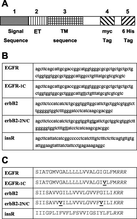
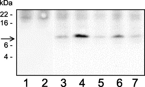
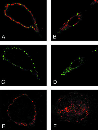
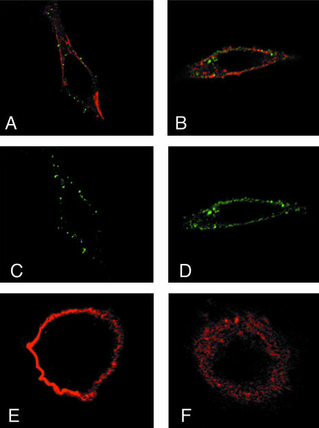
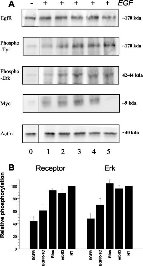
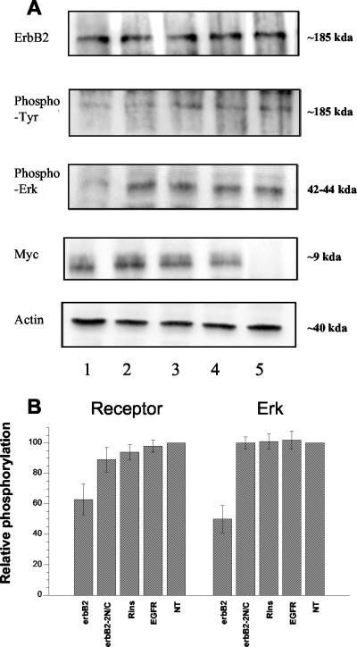
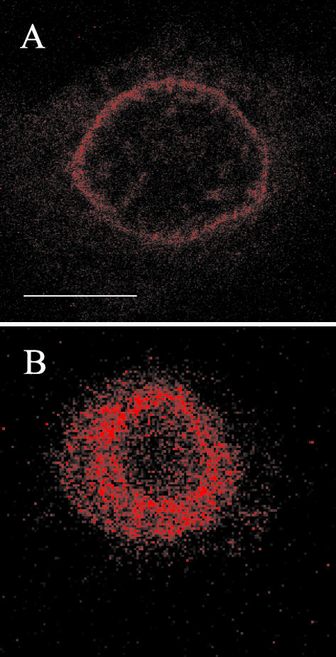
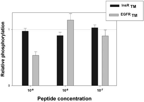
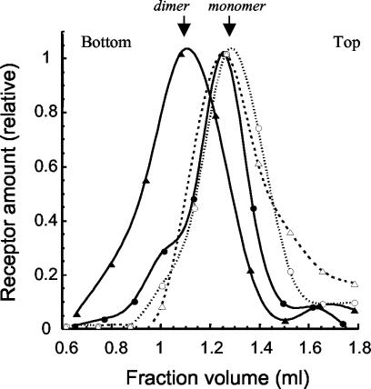
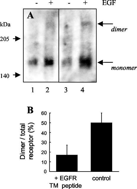
Receptor tyrosine kinases have a single transmembrane (TM) segment that is usually assumed to play a passive role in ligand-induced dimerization and activation of the receptor. However, mutations within some of these receptors, and recent studies with the epidermal growth factor (EGF) and ErbB2 receptors have indicated that interactions between TM domains do contribute to stabilization of ligand-independent and/or ligand-induced receptor dimerization and activation. One consequence of the importance of these interactions is that short hydrophobic peptides corresponding to these domains should act as specific inhibitors. To test this hypothesis, we constructed expression vectors encoding short fusion peptides encompassing native or mutated TM domains of the EGF, ErbB2, and insulin receptors. In human cell lines overexpressing the wild-type EGF receptor or ErbB2, we observed that the peptides are expressed at the cell surface and that they inhibit specifically the autophosphorylation and signaling pathway of their cognate receptor. Identical results were obtained with peptides chemically synthesized. Mechanism of action involves inhibition of dimerization of the receptors as shown by the lack of effects of mutant nondimerizing sequences, completed by density centrifugation and covalent cross-linking experiments. Our findings stress the role of TM domain interactions in ErbB receptor function, and possibly for other single-spanning membrane proteins.


Figures

Figure 1.
Figure 1.
Schematic diagram of minigenes used to encode the TM sequences. (A) Each box represents a section of the constructions made within the Invitrogen pSecTag A plasmid. (B) Oligonucleotide sequences used for cloning are indicated. (C) Amino acid sequence was as follows, using one-letter codes: signal sequence peptide (METDTLLLWVLLLWVPGSTG, 1-20), artificial extracellular sequence (extracellular tag, ET) from the multiple cloning site (DAAQPARRAVRSF, 21-33), different TM regions as indicated, including the stop transfer tribasic sequence (as indicated, 34-60; except for the EGF receptor sequence that is one amino acid longer), followed by the intracellular domain (HPAQWRPLESRGPEQKLISEEDLNSAVDHHHHHH, 61-94) containing the myc and 6His tags. Mutations introduced to decrease peptide dimerization are underlined in each sequence (see DISCUSSION), and the C-terminal tribasic stop sequence is italicized.
Figure 2.
Figure 2.
Western blotting analysis of recombinant peptide expression in A431 cells. A431 cells were transfected with pSecTag plasmids encoding for the TM domain of EGFR (column 3), ErbB2 TM (column 4), insulin receptor TM (column 5), a mutant EGFR TM (EGFR-1C, column 6), a mutant ErbB2 TM (ErbB2-2N/C, column 7), or without plasmid (columns 1 and 2). Cells were lysed in detergent 2 d after transfection and solubilized proteins were submitted to PAGE-electrophoresis and Western blotting with anti-myc tag antibodies. The arrow shows the localization of an ∼9-kDa peptide. Apparent molecular masses of protein standard are indicated at left (SeeBlue Plus2; Invitrogen). The figure represents one of several similar experiments. Identical results were obtained in other cell types, namely, SK-OV 3 as well as CHO cells.
Figure 3.
Figure 3.
Immunofluorescence analysis of whole A431 cells expressing the TM construct by using anti-EGFR and anti-ET tag antibodies. Staining of intact (A, C, and E) and permeabilized (B, D, and F) cells with anti-ET (antiexternal tag, green; A-D) and/or anti-EGFR (red; A, B, E, and F). A and C show intense membrane staining, indicating expression of the TM peptides at the cell surface, together with the receptors. B and D show membrane and vesicular labeling for both peptide and EGFR in permeabilized cells. E and F depict receptor localization in nontransfected cells.
Figure 4.
Figure 4.
Immunofluorescence analysis of whole SK-OV3 cells expressing the TM construct by using anti-ErbB2 and anti-ET tag antibodies. Staining of intact (A, C, and E) and permeabilized (B, D, and F) cells with anti-ET (antiexternal tag, green; A-D) and/or anti-ErbB2 (red; A and B, E and F). A and C show intense membrane staining, indicating expression of the TM peptides at the cell surface, together with the receptors. B and D show membrane and vesicular labeling for both peptide and ErbB2 in permeabilized cells. E and F depict receptor localization in nontransfected cells.
Figure 5.
Figure 5.
Expression, autophosphorylation, and signaling of EGF receptors in transfected A431 cells. Experiments were performed at day 2 after transfection with pSecTag plasmids encoding for the TM domain of EGFR (column 1), a mutant EGFR TM (EGFR-1C, column 2), insulin receptor TM (column 3), ErbB2 TM (column 4) or without plasmid (column 5). Column 0 represents the phosphorylation status of non-EGF-stimulated cells. Identical protein amounts of A431 cell lysates were subjected to SDS-PAGE and Western blotting with the indicated antibodies. (A) Results of one of four similar experiments. (B) Combined results after computer analysis of the blots, as histograms (mean ± SEM), for EGFR (left) and ERK 1/2 (right) phosphorylation. Values in nontransfected cells are taken as 100%.
Figure 6.
Figure 6.
Expression, autophosphorylation, and signaling of ErbB2 receptors in transfected SK-OV3 cells. Experiments were performed at day 2 after transfection with pSecTag plasmids encoding for the TM domain of ErbB2 (column 1), a mutant ErbB2 TM (ErbB2-2N/C, column 2), insulin receptor TM (column 3), EGFR TM (column 4), or without plasmid (column 5). Identical protein amounts of SK-OV3 cell lysates were subjected to SDS-PAGE and Western blotting with the indicated antibodies. (A) Results of one of four similar experiments. (B) Combined results after computer analysis of the blots, as histograms (mean ± SEM), for ErbB2 (left) and ERK 1/2 (right) phosphorylation. Values in nontransfected cells are taken as 100%.
Figure 7.
Figure 7.
Confocal fluorescence microscopy of cells in the presence of rhodamine-labeled EGFR TM peptide (A), or a glycine-rhodamine conjugate (B). Live A431 cells were grown in Lab-Tek chambers, incubated in the presence of rhodamine peptide (5 × 10⁻⁷ M) for 40 min, or glycine-rhodamine (3.5 × 10⁻⁷ M) for 3 min at 20°C before images were acquired (40× lens). White bar (A), 10 μm.
Figure 8.
Figure 8.
Autophosphorylation of EGF receptors in A431 cells incubated with synthetic TM peptides maintained in solution in detergent. Peptides corresponding to the hydrophobic core of the TM domains of EGF (gray areas) and insulin (black areas) receptors, were chemically synthesized and purified, incorporated in detergent micelles, and administered to A431 cells by supplementation of the incubation medium for 4 h. After 5 min of incubation in the presence of 10-8 M EGF, cells were lysed, and equal amounts of protein were submitted to SDS-PAGE and Western blot with anti-EGFR and anti-phosphotyrosine antibodies. Densitometric analysis of the blots was performed, and results were normalized according to the amount of immunoreactive EGFR in each sample, and expressed as percentage of the observed autophosphorylation in the absence of peptides (taken as 100%). The figure represents the results (mean ± SEM) of four similar experiments.
Figure 9.
Figure 9.
Sucrose gradient analysis of EGF receptor dimerization in the presence or absence of TM peptide. A431 cells were transfected or not with the pSecTag plasmid encoding for the EGFR TM peptide. At day 2 after transfection, cells were stimulated or not with EGF (10-8 M, 5 min), lysed with neutral detergent, and solubilized proteins were layered on top of a sucrose gradient. After centrifugation, fractions were separated and submitted to SDS-PAGE electrophoresis and Western blotting with anti-EGF receptor antibodies. The figure shows the densitometric quantification of a typical blot out of three similar experiments. Values were normalized relatively to the highest peak, to account for differences in total receptor content. Symbols are as follow: triangles, untransfected A431 cells; circles, cells transfected with the pSecTag plasmid encoding for the EGFR TM peptide. Open symbols and dotted lines are for unstimulated cells; filled symbols and full lines are for cells stimulated with EGF. Top and bottom of the gradient are indicated.
Figure 10.
Figure 10.
Analysis of EGF receptor dimerization in the presence or absence of TM peptide by using a cross-linking agent. A431 cells were transfected or not with the pSecTag plasmid encoding for the EGFR TM peptide. At day 2 after transfection, cells were stimulated or not with EGF (10-8 M, 5 min), cross-linked with disuccinimidyl suberate, and lysed. Samples were immunoprecipitated and submitted to SDS-PAGE electrophoresis and Western blotting with anti-EGF receptor antibodies. (A) Typical blot out of four experiments. The first two columns represent cells transfected with the EGFR TM peptide without (-) and with (+) stimulation with EGF. Columns 3 and 4 represent control untransfected cells. (B) Mean ratio (± SEM) of dimer versus total receptor (dimer/monomer + dimer) in EGF-stimulated cells after densitometric analysis of the blots.

References

    1. Bargmann, C.I., Hung, M.C., and Weinberg, R.A. (1986). Multiple independent activations of the neu oncogene by a point mutation altering the transmembrane domain of p185. Cell 45, 649-657. - PubMed
    1. Bell, C.A., Tynan, J.A., Hart, K.C., Meyer, A.N., Robertson, S.C., and Donoghue, D.J. (2000). Rotational coupling of the transmembrane and kinase domains of the Neu receptor tyrosine kinase. Mol. Biol. Cell 11, 3589-3599. - PMC - PubMed
    1. Blume-Jensen, P., and Hunter, T. (2001). Oncogenic kinase signalling. Nature 411, 355-365. - PubMed
    1. Burke, C.L., Lemmon, M.A., Coren, B.A., Engelman, D.M., and Stern, D.F. (1997). Dimerization of the p185neu transmembrane domain is necessary but not sufficient for transformation. Oncogene 14, 687-696. - PubMed
    1. Cheatham, B., Shoelson, S.E., Yamada, K., Goncalves, E., and Kahn, C.R. (1993). Substitution of the erbB-2 oncoprotein transmembrane domain activates the insulin receptor and modulates the action of insulin and insulin-receptor substrate 1. Proc. Natl. Acad. Sci. USA 90, 7336-7340. - PMC - PubMed

Publication types