. 2004 Jul;15(7):3333-44.
doi: 10.1091/mbc.e03-12-0878. Epub 2004 May 14.

The entire Nup107-160 complex, including three new members, is targeted as one entity to kinetochores in mitosis

Affiliations

The entire Nup107-160 complex, including three new members, is targeted as one entity to kinetochores in mitosis

Isabelle Loïodice et al. Mol Biol Cell. 2004 Jul.

Abstract

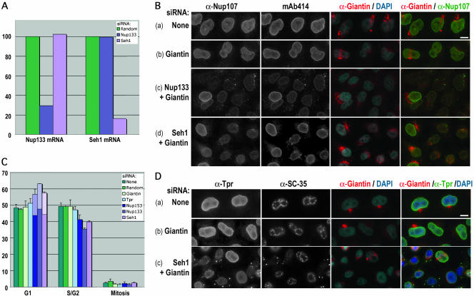
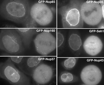
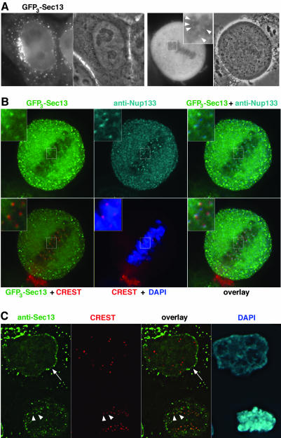
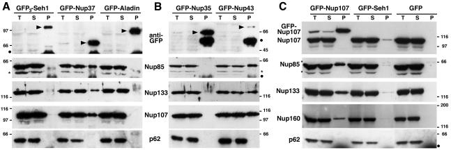
In eukaryotes, bidirectional transport of macromolecules between the cytoplasm and the nucleus occurs through elaborate supramolecular structures embedded in the nuclear envelope, the nuclear pore complexes (NPCs). NPCs are composed of multiple copies of approximately 30 different proteins termed nucleoporins, of which several can be biochemically isolated as subcomplexes. One such building block of the NPC, termed the Nup107-160 complex in vertebrates, was so far demonstrated to be composed of six different nucleoporins. Here, we identify three WD (Trp-Asp)-repeat nucleoporins as new members of this complex, two of which, Nup37 and Nup43, are specific to higher eukaryotes. The third new member Seh1 is more loosely associated with the Nup107-160 complex biochemically, but its depletion by RNA interference leads to phenotypes similar to knock down of other constituents of this complex. By combining green fluorescent protein-tagged nucleoporins and specific antibodies, we show that all the constituents of this complex, including Nup37, Nup43, Seh1, and Sec13, are targeted to kinetochores from prophase to anaphase of mitosis. Together, our results indicate that the entire Nup107-160 complex, which comprises nearly one-third of the so-far identified nucleoporins, specifically localizes to kinetochores in mitosis.


Figures

Figure 4.
Figure 4.
Effects of Seh1 depletion in HeLa cells are reminiscent to those observed upon Nup133 depletion. (A) Quantitative RT-PCR analysis of Seh1 and Nup133 mRNAs from HeLa cells treated for 3 d with siRNA duplexes specific for Seh1 or Nup133 or a random siRNA. The amounts of Seh1 and Nup133 mRNAs were arbitrarily set at 100% for cells treated with the random siRNA. (B) Widefield microscopy images of immunofluorescence of HeLa cells treated for 3 d with no siRNA (a), Giantin siRNA duplexes (b), or Giantin siRNA duplexes together with siRNA duplexes specific for Nup133 (c) or Seh1 (d) and labeled with anti-Nup107, mAb414, and anti-Giantin antibodies. DNA was stained with 4,6-diamidino-2-phenylindole (DAPI). Note in c and d the wild-type labeling of Nup107 and mAb414 in cotransfected cells that are not depleted for Giantin. As previously observed in Nup107-depleted cells (Walther et al., 2003a), cytoplasmic foci are slightly less abundant in Seh1-depleted cells compared with Nup133-depleted cells. (C) Cell cycle analyses of HeLa cells treated for 3 d with no siRNA, a random siRNA, or with siRNA duplexes specific for Tpr, Nup153, Nup133, or Seh1. S/G2 cells were detected by BrdU and cyclin B1 staining, and mitotic cells by phase contrast. The clear part of the histograms corresponds to the percentage of cells with SC-35 cytoplasmic foci. Standard deviations are indicated. (D) Widefield microscopy images of immunofluorescence of HeLa cells treated for 3 d with no siRNA (a), Giantin siRNA duplexes (b), or Giantin siRNA duplexes (c) together with siRNA duplexes specific for Seh1. Cells were labeled with anti-Tpr, anti-SC-35, anti-Giantin, and DAPI. Bars, 10 μm.
Figure 5.
Figure 5.
Constituents of the Nup107-160 complex, but not GFP-Nup35, are targeted to kinetochores. Widefield microscopy images of live interphase and mitotic HeLa cells expressing, as indicated, GFP-Nup85, GFP-Nup35, GFP-Nup160, GFP-Seh1, GFP-Nup37, or GFP-Nup43. Whereas all these GFP-tagged nucleoporins are targeted to the nuclear envelope of interphase cells, fluorescent foci can be visualized at the metaphase plate of mitotic cells expressing these various fusions, with the exception of GFP-Nup35-expressing cells.
Figure 8.
Figure 8.
A fraction of Sec13 localizes to both the nuclear envelope and kinetochores. (A) Widefield microscopy images of live interphase and mitotic HeLa cells expressing GFP3-Sec13. The fluorescent signal and the corresponding phase contrast images are shown. The inset shows a threefold enlargement of the marked area within the metaphase plate, where bright GFP3-Sec13 foci can be detected (arrowheads). (B) Deconvolved images of GFP3-Sec13-expressing cells double labeled with affinity-purified anti-Nup133 antibody and the CREST serum. DNA was stained with DAPI. Insets show a threefold enlargement of the marked area. Note the overlap between the GFP3-Sec13 and the anti-Nup133 stainings that are more peripheral compared with the CREST labeling. (C) Deconvolved images of HeLa cells double labeled with affinity-purified anti-Sec13 antibody and the CREST serum. DNA was stained with DAPI. The arrow on the interphase cell (top) points to the nuclear envelope, labeled with the Sec13 antibody, and arrowheads indicate the position of kinetochores, labeled with both the anti-Sec13 antibody and CREST serum in an anaphase cell (bottom).
Figure 1.
Figure 1.
Immunoprecipitation of soluble extracts from HeLa cells expressing GFP3-Seh1, GFP-Nup37, GFP-Aladin (A); GFP-Nup35, GFP-Nup43 (B); and GFP-Nup107, GFP-Seh1, or GFP (C) by using anti-GFP antibodies. Equivalent amounts of total extracts (T) and immune supernatants (S) and tenfold equivalents of the immune pellets (P) were analyzed by immunoblot by using, as indicated, anti-GFP, Nup85, Nup133, Nup107, or Nup160 antibodies or the mAb414 antibody that mainly recognizes p62. Arrowheads in A and B point to the position of the GFP fusions. Dots indicate IgG heavy chains and stars a protein that cross-reacts with the anti-Nup85 antibody. Molecular mass markers are in kilodaltons.
Figure 3.
Figure 3.
GFP-Nup43 and GFP3-Sec13 coimmunoprecipitate with myc6-Nup37 and the other constituents of the Nup107-160 complex. Immunoprecipitation using anti-GFP antibodies of soluble extracts from HeLa cells coexpressing GFP-Nup35, GFP-Nup43, or GFP3-Sec13, and myc6-Nup37. Equivalent amounts of total extracts (T) and immune supernatants (S) and tenfold equivalents of the immune pellets (P) were analyzed by immunoblot by using anti-GFP, anti-myc (that recognizes the myc6-Nup37 fusion), Nup85, Nup160, Nup133, Nup96, Nup107, Nup85, or Sec31 antibodies. Arrowheads indicate the position of the GFP fusions. Molecular mass markers on the left are in kilodlatons.
Figure 2.
Figure 2.
Analysis of myc6-Nup37 and myc6-Seh1 behavior by sucrose gradients. (A) Soluble extracts from HeLa cells (b and f) or from HeLa cells expressing myc6-Nup37 (a and d) or myc6-Seh1 (c and e) were loaded on continuous 20-35% sucrose gradients and centrifuged at 100,000 × g for 16 h. Fractions from the top (fraction 1) to the bottom (fraction 26) of the gradient were analyzed by Western blot by using anti-Nup133 (a), Nup85 (b), Nup37 (c), or myc (d and e) antibodies, or the mAb414 antibody (f). (B) Fractions 11-14 from the above-described sucrose gradient (b) were pooled, and 50 μl of the resulting sample was loaded on a Superose 6 (PC 3.2/30) gel filtration chromatography column. The initial sample (L) and the Superose 6 fractions were analyzed by Western blot by using anti-Nup133, Nup85, and Nup37 antibodies. Molecular size markers used to calibrate the sucrose gradient and the Superose 6 column are indicated. Thick bars denote the position of the Nup107-160 complex.
Figure 6.
Figure 6.
GFP-Nup37 is localized at kinetochores from prophase to anaphase. (A) Deconvolved images of fixed metaphase HeLa cells expressing GFP-Nup37 (shown in green) and labeled with anti-CENP-F antibody (shown in red) and the CREST serum (shown in blue). Insets showing enlargements of the marked area reveal that GFP-Nup37 lies distal to the centromeres labeled with the CREST serum, and slightly more central than CENP-F, an outer kinetochore protein. (B) Fixed HeLa cells expressing GFP-Nup37 were labeled with the CREST serum and DAPI. Deconvolved images of interphase (a), prophase (b), late prophase/prometaphase (c), early anaphase (d), and late anaphase (e) are presented. The insets show enlargements of the marked area. Arrows point to the position of centromeres. Note the weak kinetochore labeling of GFP-Nup37 in early prophase and the persistence of this staining until late anaphase.
Figure 7.
Figure 7.
Nup96 stably interacts with the Nup107-160 complex in mitosis and is localized at kinetochores in mitotic HeLa cells. (A) Immunoprecipitation of interphase (I) or mitotic (M) HeLa S3 cell extracts by using affinity-purified anti-Nup133 antibodies. Equivalent amounts of total extracts (T) and depleted supernatants (S) and fivefold equivalents of the immune pellets (P) were analyzed by immunoblot by using antibodies directed against Nup133, Nup96, Nup85, Nup37, and Sec13. Similar coimmunoprecipitation efficiencies were obtained with these various nucleoporins from both interphase and mitotic extracts. Note the very efficient codepletion and coprecipitation of Nup96 together with Nup133. (B) Deconvolved images of HeLa cells double-labeled with affinity-purified anti-Nup96 and monoclonal anti-CENP-F antibodies. DNA was stained with DAPI. Note that Nup96 is clearly detected at kinetochores in prophase (cell on the left) and metaphase.

References

    1. Allen, N.P.C., Patel, S.S., Huang, L., Chalkley, R.J., Burlingame, A., Lutzmann, M., Hurt, E.C., and Rexach, M. (2002). Deciphering networks of protein interactions at the nuclear pore complex. Mol. Cell. Proteomics 1, 930-946. - PubMed
    1. Askjaer, P., Galy, V., Hannak, E., and Mattaj, I.W. (2002). Ran GTPase cycle and importins alpha and beta are essential for spindle formation and nuclear envelope assembly in living Caenorhabditis elegans Embryos. Mol. Biol. Cell 13, 4355-4370. - PMC - PubMed
    1. Babu, J.R., Jeganathan, K.B., Baker, D.J., Wu, X., Kang-Decker, N., and Van Deursen, J.M. (2003). Rae1 is an essential mitotic checkpoint regulator that cooperates with Bub3 to prevent chromosome missegregation. J. Cell Biol. 160, 341-353. - PMC - PubMed
    1. Belgareh, N., et al. (2001). An evolutionarily conserved NPC subcomplex, which redistributes in part to kinetochores in mammalian cells. J. Cell Biol. 154, 1147-1160. - PMC - PubMed
    1. Boehmer, T., Enninga, J., Dales, S., Blobel, G., and Zhong, H. (2003). Depletion of a single nucleoporin, Nup107, prevents the assembly of a subset of nucleoporins into the nuclear pore complex. Proc. Natl. Acad. Sci. USA 100, 981-985. - PMC - PubMed

Publication types

Substances