Comparative Study
. 2004 Jun;10(6):954-64.
doi: 10.1261/rna.7030704.

The structure of a ribosomal protein S8/spc operon mRNA complex

Affiliations
Comparative Study

The structure of a ribosomal protein S8/spc operon mRNA complex

Helen J Merianos et al. RNA. 2004 Jun.

Abstract

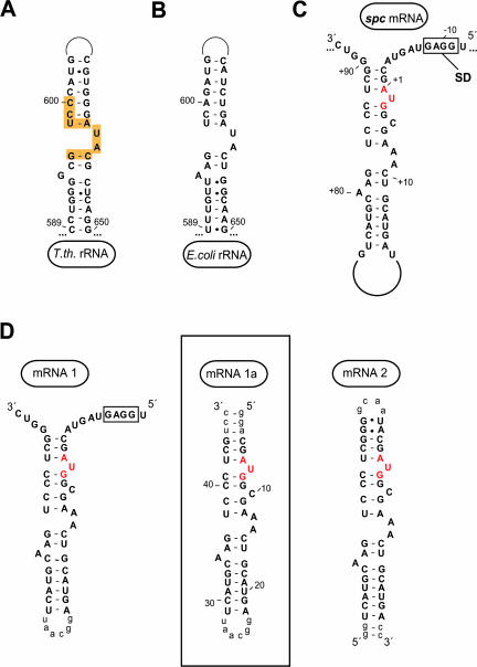
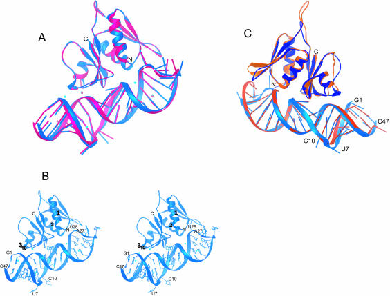
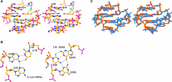
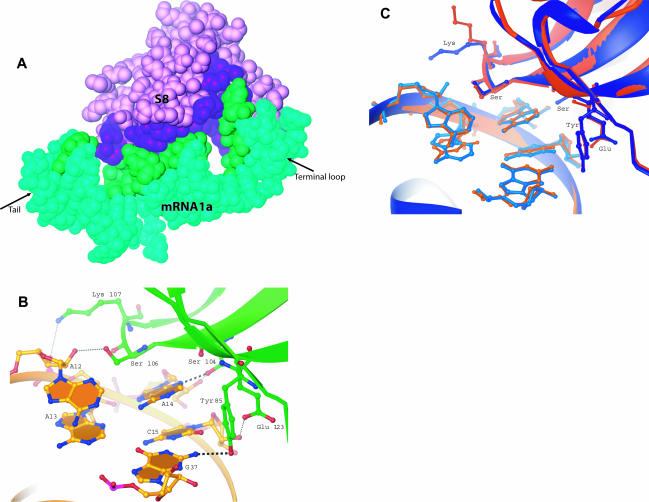
In bacteria, translation of all the ribosomal protein cistrons in the spc operon mRNA is repressed by the binding of the product of one of them, S8, to an internal sequence at the 5' end of the L5 cistron. The way in which the first two genes of the spc operon are regulated, retroregulation, is mechanistically distinct from translational repression by S8 of the genes from L5 onward. A 2.8 A resolution crystal structure has been obtained of Escherichia coli S8 bound to this site. Despite sequence differences, the structure of this complex is almost identical to that of the S8/helix 21 complex seen in the small ribosomal subunit, consistent with the hypothesis that autogenous regulation of ribosomal protein synthesis results from conformational similarities between mRNAs and rRNAs. S8 binding must repress the translation of its own mRNA by inhibiting the formation of a ribosomal initiation complex at the start of the L5 cistron.


Figures

FIGURE 1.
FIGURE 1.
Secondary structure of RNA molecules to which ribosomal protein S8 binds. (A,B) Helix 21 of 16S rRNA from T. thermophilus and E. coli, respectively. Yellow regions indicate S8 contacts within the T. thermophilus 30S (Brodersen et al. 2002). (C) Wild-type regulatory site in the spc operon mRNA from E. coli. The start codon for the L5 gene is shown in red, and the Shine-Dalgarno region is boxed. (D) mRNA constructs used in this study. Lowercase nucleotides were added for stability or transcriptional purposes and are not part of the wild-type sequence. The boxed construct was crystallized in complex with E. coli S8.
FIGURE 2.
FIGURE 2.
Typical electron-density. Stereoview depicting typical electron density from a composite omit map contoured to 1.0σ at a resolution limit of 2.7 Å. The region of mRNA1a shown is a portion of the S8-binding site.
FIGURE 3.
FIGURE 3.
The structure of E. coli S8/mRNA1a and its comparison with the T. thermophilus S8/helix 21 structure. (A) By using the phosphorus atoms of the S8-binding site (A12–G17/C34–U38) and the Cαs of S8, Complex A (blue) was superimposed on complex B (pink). (B) Stereoview of the structure of the E. coli S8–mRNA complex shown in blue (complex A), with the Zn2+ ions shown in cyan. (C) A comparison of E. coli complex A (blue) with T. thermophilus S8/helix 21 (orange; Wimberly et al 2000). Superposition was done by using the phosphorus atoms of the S8-binding site (A12–G17/C34–U38) and the Cα atoms of S8. Note that the complexes have been rotated 180° relative to those shown in A and B.
FIGURE 4.
FIGURE 4.
Structure of S8s mRNA and rRNA-binding sites. (A) Stereoview of E. coli mRNA-binding site (A12–G17/C34–U38). Zn ions, shown in cyan, are tetrahedrally coordinated and participate in water-mediated interactions with the mRNA. (B) The U16–A35–A36 base triple from E. coli mRNA1a (left) and the corresponding base triple from T. thermophilus G644–C596–G595 (right). (C) Stereoview of the superposition of the E. coli mRNA-binding site (blue) with the T. thermophilus rRNA-binding site (orange). The C1′ atoms of the conserved residues, which interact with S8 (C15, G37, A14, A12), were used for the superposition.
FIGURE 5.
FIGURE 5.
S8–RNA interactions. (A) Surface contacts between E. coli S8 (purple) and mRNA1a (green). Buried surface area on the mRNA is shown in dark green and buried surface from S8 is shown in dark purple. (B) Hydrogen bonding between E. coli S8 and its spc operon mRNA-binding site. Two base-specific hydrogen bonds occur (Y85:G37, and S104:A14). All other protein hydrogen bonds involve phosphates or sugars. (C) A comparison of RNA–protein interactions between E. coli S8/mRNA (blue) and T. thermophilus S8/rRNA (orange). Phosphorus atoms and Cα atoms shown were used for the superposition.

References

    1. Brodersen, D.E., Clemons Jr., W.M., Carter, A.P., Wimberly, B.T., and Ramakrishnan, V. 2002. Crystal structure of the 30S ribosomal subunit from Thermus thermophilus: Structure of the proteins and their interactions with 16S RNA. J. Mol. Biol. 316: 725–768. - PubMed
    1. Brunger, A.T., Adams, P.D., Clore, G.M., DeLano, W.L., Gros, P., Grosse-Kunstleve, R.W., Jiang, J.S., Kuszewski, J., Nilges, N., Pannu, N.S., et al. 1998. Crystallography and NMR system: A new software suite for macromolecular structure determination. Acta Crystallogr. D Biol. Crystallogr. 54: 905–921. - PubMed
    1. Carson, M. 1991. Ribbons 2.0. J. Appl. Crystallogr. 24: 958–961.
    1. Cerretti, D.P., Mattheakis, L.C., Kearney, K.R., Vu, L., and Nomura, M. 1988. Translational regulation of the spc operon in Escherichia coli: Identification and structural analysis of the target site for S8 repressor protein. J. Mol. Biol. 204: 309–329. - PubMed
    1. Chothia, C. and Janin, J. 1975. Principles of protein–protein recognition. Nature 256: 705–708. - PubMed

Publication types

MeSH terms

Associated data

LinkOut - more resources