. 2004 Jun;10(6):965-77.
doi: 10.1261/rna.7090104.

Functions for S. cerevisiae Swd2p in 3' end formation of specific mRNAs and snoRNAs and global histone 3 lysine 4 methylation

Affiliations

Functions for S. cerevisiae Swd2p in 3' end formation of specific mRNAs and snoRNAs and global histone 3 lysine 4 methylation

Bernhard Dichtl et al. RNA. 2004 Jun.

Abstract

The Saccharomyces cerevisiae WD-40 repeat protein Swd2p associates with two functionally distinct multiprotein complexes: the cleavage and polyadenylation factor (CPF) that is involved in pre-mRNA and snoRNA 3' end formation and the SET1 complex (SET1C) that methylates histone 3 lysine 4. Based on bioinformatic analysis we predict a seven-bladed beta-propeller structure for Swd2p proteins. Northern, transcriptional run-on and in vitro 3' end cleavage analyses suggest that temperature sensitive swd2 strains were defective in 3' end formation of specific mRNAs and snoRNAs. Protein-protein interaction studies support a role for Swd2p in the assembly of 3' end formation complexes. Furthermore, histone 3 lysine 4 di-and tri-methylation were adversely affected and telomeres were shortened in swd2 mutants. Underaccumulation of the Set1p methyltransferase accounts for the observed loss of SET1C activity and suggests a requirement for Swd2p for the stability or assembly of this complex. We also provide evidence that the roles of Swd2p as component of CPF and SET1C are functionally independent. Taken together, our results establish a dual requirement for Swd2p in 3' end formation and histone tail modification.


Figures

FIGURE 1.
FIGURE 1.
Swd2p carries seven WD repeats and is conserved within eukaryotes. (A) Multiple sequence alignment of a representative set of the Swd2 family. The sequences are: Ag_CG51024: Anopheles gambiae (tr:Q7Q1N9); Dm_CG17293 and Dm_CG3515: Drosophila melanogaster (tr:Q9VLN1 and Q9VQD1); Mm_9430077D24Rik: Mus musculus (tr:Q8BFQ4); Hs_UNQ9342: Homo sapiens (trnew:AAQ88631); Zf_Q803V6: Danio rerio (Q803V6); Sp_Swd2.1 and Sp_Swd2.2: S. pombe (tr:O60137 and tr:Q9UT39); Sc_Swd2: S. cerevisiae (sw:P36104); Nc_NCU07885.1: Neurospora crassa (tr:Q7SB08); Ac_Q9P423: Ajellomyces capsulata (tr:Q9P423); At_T15N1_20 and At_AT5G66240: Arabidopsis thaliana (tr:Q9LYK6 and tr:Q8RXD8); Ce_C33H5.7 and Ce_C33H5.6 Caenorhabditis elegans (tr:Q18403 and Q18404); the latter sequence was repredicted from the genomic sequence as described in Materials and Methods. The databases are: tr: trembl; trnew: trembl_new, and sw: SwissProt. Not included is an additional human sequence (GenBank: LOC131060), which is virtually identical to Hs_UNQ9342. It is not clear if these two sequences derive from the same gene. The first 43 residues of Ag_CG51024 and the last 72 residues of Dm_CG3515 are not shown. The approximate positions of seven predicted WD-40 repeats (see B) are indicated with green bars above the sequence. The darker green squares indicate the position of the sequence most likely to be the “WD” motif. (B) A profile–profile dot plot using a profile generated from the alignment in A shows six off-diagonal lines indicating that the family has seven WD-40 repeats. The plot was generated with a window of 17 and a dot-depth of 5. Note that the rulers on the axes are not identical to the ruler in A because gapped regions are excluded in the profile.
FIGURE 2.
FIGURE 2.
Swd2 is required for 3′ end formation of specific mRNAs and snoRNAs. Northern analysis of 20 μg total RNA extracted from wild-type, swd2-2, swd2-6, and ssu72-2 cells following growth in YPD medium at 25°C or after a shift to 37°C for the indicated times. Blots were produced in duplicate. (AF) Probed as indicated at the bottom of each panel; migration of RNAs is indicated on the right. In the middle of each panel a schematic presentation indicates the genomic arrangement of the analyzed genes and the relative direction of transcription.
FIGURE 3.
FIGURE 3.
Mutations in SWD2 do not interfere with 3′ end cleavage of CYC1 pre-mRNA in vitro. (A) Transcriptional run-on analysis. Slot-hybridizations and quantification of run-on transcripts obtained after transcription run-on analysis (Birse et al. 1998). Wild-type, swd2-2, and swd2-6 cells were grown in synthetic medium lacking uracil and 1 mM CuSO4 at 25°C or shifted to 37°C in a water bath for 1 h. P1 to P6 represent probes complementary to CYC1 transcripts as indicated; empty M13 and ACT1 probes served as controls. Values obtained by PhosphorImager Scanning (Storm, Molecular Dynamics) were corrected by subtraction of the M13 background signal and normalized to the value of P1 that was fixed at 100%. (B) 3′ end cleavage in vitro. Assays were performed with extracts prepared from the indicated strains. Input lanes represent mock-treated reactions. Marker bands (HpaII-digested pBR322) are indicated on the left. Internally [32P]-labeled CYC1 RNA was used as substrate. The positions of full-length RNA (CYC1), 5′ and 3′ cleavage products are indicated. Cleavage was performed either at 30°C (lanes 27) or at 37°C, following a 5-min preincubation of extract and reaction mixture at this temperature (lanes 813).
FIGURE 4.
FIGURE 4.
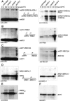
Swd2p bridges core-CPF, APT, and CF IA subcomplexes of the 3′ end formation machinery. (AC) Pull-down experiments with 1 μg GST or GST-Swd2p (Swd2p) and [35S]-methionine-labeled proteins as indicated. Also indicated is the association of tested subunits with core-CPF (A), the APT complex (B), or CF I (C). Input lanes show 10% of in vitro translation reactions included in binding reactions. Note that the band visible in the Pap1p lane (lane 20) does not correspond to full-length protein. (D) Schematic representation of subcomplexes and individual protein subunits of the yeast 3′ end formation machinery. Abbreviations for subcomplexes are underlined. The CF IA and holo-CPF complexes constitute most components required for 3′ end formation in vitro. Holo-CPF was suggested to contain core-CPF (previously called PF I) and APT subcomplexes (Nedea et al. 2003). Core-CPF includes the smaller CF II complex as indicated by a dashed line. Polypeptides interacting with GST-Swd2p in vitro are encircled and indicated by arrows.
FIGURE 5.
FIGURE 5.
Swd2p is required for SET1C activity. (A) Protein lysates were produced from wild-type (wt), swd2, ΔSET1, K4A, and K9A mutant strains as indicated on top of each lane. The wild-type and swd2 mutant strains were grown in YPD medium at 25°C or transferred for 4 h to a 37°C water bath. ΔSET1, K4A, and K9A strains were grown at 30°C. Following Western transfer filters were probed with polyclonal antisera directed against the indicated proteins. Asterisks mark cross-reacting bands that served as control for equal loading. (B) Southern blot of genomic DNA obtained from the indicated strains. swd2 mutants and the isogenic wild-type strain were grown in YPD medium at 25°C (lanes 14) or transferred to a 37°C water bath for 8 h (lanes 58). Ten micrograms genomic DNA were digested with XhoI and resolved on 1% agarose gels. Southern hybridization was performed with a telomere specific probe. Migration of telomere fragments is indicated on the right. (C) Western analysis of extracts from strains expressing ProtA fusions to wild-type and mutant Swd2p proteins. The indicated strains were grown at 25°C or shifted to 37°C for 4 h in YPD medium. Fifty micrograms total protein were analyzed by Western with specific antibodies. ProtA-Swd2p was detected with a polyclonal IgG. Brackets on the right indicate the association of proteins with SET1C or CPF. (D) Northern analysis as described in the legend of Figure 2 ▶. Total RNA was extracted from the strains indicated on top of each lane following growth at 30°C in YPD medium. Note that this temperature is semipermissive for swd2-2 and swd2-6 strains. Probes were as described in the legend of Figure 2 ▶ and are indicated below each panel. 18S rRNA served as loading control and was detected by a 5′ end-labeled oligonucleotide. The detected RNA species are indicated on the right of the panels.
FIGURE 6.
FIGURE 6.
Swd2p associates with separable high molecular weight complexes. Protein extracts were produced from wild-type (wt; ProtA-Swd2p) and mutant (mut; ProtA-swd2-2p) strains following growth at 25°C or after a shift to 37°C for 4 h as indicated. Protein was separated by SMART Superdex 200 gel filtration and analyzed by Western blotting. Fraction numbers and input material are indicated on the top of the panels and the migration of molecular weight markers separated under the same conditions is indicated at the bottom of the panels. Also indicated is the migration of peak fractions of Set1p protein (SET1C peak) and of Ysh1p protein (CPF peak). On the right of the panels the detected proteins are indicated.

References

    1. Alen, C., Kent, N.A., Jones, H.S., O’Sullivan, J., Aranda, A., and Proudfoot, N.J. 2002. A role for chromatin remodeling in transcriptional termination by RNA polymerase II. Mol. Cell 10: 1441–1452. - PubMed
    1. Altschul, S.F., Madden, T.L., Schaffer, A.A., Zhang, J., Zhang, Z., Miller, W., and Lipman, D.J. 1997. Gapped BLAST and PSI-BLAST: A new generation of protein database search programs. Nucleic Acids Res. 25: 3389–3402. - PMC - PubMed
    1. Apweiler, R., Bairoch, A., Wu, C.H., Barker, W.C., Boeckmann, B., Ferro, S., Gasteiger, E., Huang, H., Lopez, R., Magrane, M., et al. 2004. UniProt: The universal protein knowledgebase. Nucleic Acids Res. 32: 115–119. - PMC - PubMed
    1. Barabino, S.L.M., Ohnacker, M., and Keller, W. 2000. Distinct roles of two Yth1p domains in 3′-end cleavage and polyadenylation of yeast pre-mRNAs. EMBO J. 19: 3778–3787. - PMC - PubMed
    1. Bentley, D. 2002. The mRNA assembly line: Transcription and processing machines in the same factory. Curr. Opin. Cell. Biol. 14: 336–342. - PubMed

Publication types

MeSH terms