. 2004 May;113(10):1390-7.
doi: 10.1172/JCI20402.

Induction of B7-1 in podocytes is associated with nephrotic syndrome

Affiliations

Induction of B7-1 in podocytes is associated with nephrotic syndrome

Jochen Reiser et al. J Clin Invest. 2004 May.

Abstract

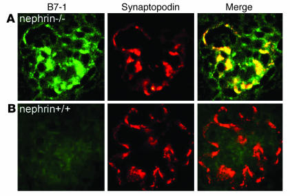
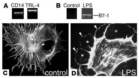
Kidney podocytes and their slit diaphragms form the final barrier to urinary protein loss. This explains why podocyte injury is typically associated with nephrotic syndrome. The present study uncovered an unanticipated novel role for costimulatory molecule B7-1 in podocytes as an inducible modifier of glomerular permselectivity. B7-1 in podocytes was found in genetic, drug-induced, immune-mediated, and bacterial toxin-induced experimental kidney diseases with nephrotic syndrome. The clinical significance of our results is underscored by the observation that podocyte expression of B7-1 correlated with the severity of human lupus nephritis. In vivo, exposure to low-dose LPS rapidly upregulates B7-1 in podocytes of WT and SCID mice, leading to nephrotic-range proteinuria. Mice lacking B7-1 are protected from LPS-induced nephrotic syndrome, suggesting a link between podocyte B7-1 expression and proteinuria. LPS signaling through toll-like receptor-4 reorganized the podocyte actin cytoskeleton in vitro, and activation of B7-1 in cultured podocytes led to reorganization of vital slit diaphragm proteins. In summary, upregulation of B7-1 in podocytes may contribute to the pathogenesis of proteinuria by disrupting the glomerular filter and provides a novel molecular target to tackle proteinuric kidney diseases. Our findings suggest a novel function for B7-1 in danger signaling by nonimmune cells.


Figures

Figure 1
Figure 1
B7-1 expression in podocytes. (A) Differential display PCR of WT (+/+) and α3_/_ (_/_) podocytes. The arrow indicates the position of B7-1. (B) Induction of B7-1 mRNA expression in α3_/_ podocytes. RT-PCR from three independent samples (a_c) on serial dilution (1:5 to 1:25) of cDNA standardized for β-actin. Con, H2O control. (C) Rescue (Res) of α3_/_ podocytes by stable transfection with human α3 integrin restored B7-1 expression to WT levels. RT-PCR for GAPDH shows equal RNA concentrations. (D) Dose-dependent increase of B7-1 mRNA in WT podocytes in response to puromycin (0.1, 1, 5, 10, and 50 ∝g/ml). (E) Time course of B7-1 and α3 integrin mRNA expression in puromycin aminonucleoside_treated (PAN-treated) WT podocytes. As early as 6 and 24 hours, B7-1 is induced by PAN, whereas α3 integrin levels remain unchanged. (F) B7-1 is not expressed in E18 WT kidneys (left panel) but is expressed in podocytes of α3_/_ kidneys as shown by double labeling with synaptopodin (right panels).
Figure 2
Figure 2
B7-1 expression in podocytes in murine and human lupus nephritis correlates with the severity of proteinuria. (A) B7-1 is not expressed in normal mouse kidney (left). In the lupus kidney, strong glomerular (G) B7-1 expression is observed (middle). In addition, infiltrating interstitial B cells stain positive (arrows). At higher magnification (right), B7-1 expression is found in podocytes (arrows) in the glomerulus. (B) Double labeling with synaptopodin confirmed that intraglomerular B7-1 expression is restricted to podocytes in 4+ proteinuric (4+ prot) nephritis. (C) B7-1 is absent in an age-matched lupus mouse with trace proteinuria (trace prot). (D and E) In human lupus nephritis, glomerular B7-1 expression increases from WHO class II to V and colocalizes with synaptopodin in podocytes (E).
Figure 3
Figure 3
Induction of B7-1 in podocytes of nephrin_/_ mice. (A) B7-1 is induced in podocytes of nephrin_/_ mice as shown by double labeling with synaptopodin. (B) B7-1 is absent from podocytes of WT littermate controls. These data establish that the expression of B7-1 in podocytes in vivo can occur independently of immune complexes or inflammatory cells.
Figure 4
Figure 4
LPS-induced upregulation of B7-1 and actin cytoskeleton reorganization in podocytes in vitro. (A) Cultured podocytes express the LPS receptors CD14 and TLR-4 as shown by RT-PCR. (B) LPS treatment upregulates B7-1 mRNA expression and reorganizes the podocyte cytoskeleton from a stress fiber pattern (C), creating a cortical ring of F-actin (D; arrowheads) and a submembranous space devoid of actin fibers (asterisks).
Figure 5
Figure 5
LPS-induced FP effacement and proteinuria are B7-1 dependent. (A) LPS injection (left panel) and PBS injection (right panel) do not cause renal inflammation 24 hours after injection in WT mice. (B) LPS injection induces B7-1 in podocytes as shown by double labeling with synaptopodin. (C) PBS-injected control mice do not upregulate B7-1. (D) LPS injection causes severe FP effacement within 24 hours in WT mice (left panel; arrows) but not in PBS-injected injected control animals (right panel). (E and F) B7-1 is upregulated in podocytes of LPS-treated SCID mice (E), but not in PBS-injected SCID controls (F). (G) LPS injection causes severe proteinuria in 129, C57BL/6, and BALB/c WT mice and SCID mice but not in B7-1_/_ mice. Asterisks indicate statistically significant changes (see text for details).
Figure 6
Figure 6
Redistribution of SD proteins to sites of B7-1 engagement. (A) In untreated α3_/_ podocytes, nephrin is expressed at cell-cell contacts. (B and C) Incubation of podocytes with microbeads coated with CTLA-Ig (B) or anti_B7-1 antibody (C) redistributes nephrin around microbeads (arrows). (D) The association of nephrin with microbeads was confirmed by confocal microscopy. (E_L) Similar redistribution patterns were seen for CD2AP (E_H) and ZO-1 (I_L). Arrows in F and J: accumulation of CD2AP (F) and ZO-1 (J) around microbeads. Asterisk in K: microbead. (M) In contrast, microbeads coated with an irrelevant antibody could to some extent adhere to podocytes (arrowhead) but did not affect the localization of ZO-1 at cell-cell contacts (arrows).
Figure 7
Figure 7
Pathways leading to B7-1_mediated proteinuria. Various stimuli may lead to podocyte B7-1 induction. These include genetic stimuli (e.g., deletion of α3 integrin or nephrin), toxic stimuli (PAN-induced reactive oxygen species [ROS]), or direct stimulation of the TLR-4/CD14 receptor on the podocyte. B7-1 then induces FP effacement and disruption of the SD complex, thereby modifying glomerular permselectivity.

References

    1. Somlo S, Mundel P. Getting a foothold in nephrotic syndrome. Nat. Genet. 2000;24:333–335. - PubMed
    1. Kriz W, Gretz N, Lemley KV. Progression of glomerular diseases: is the podocyte the culprit? Kidney Int. 1998;54:687–697. - PubMed
    1. Kestila M, et al. Positionally cloned gene for a novel glomerular protein—nephrin—is mutated in congenital nephrotic syndrome. Mol. Cell. 1998;1:575–582. - PubMed
    1. Shih NY, et al. Congenital nephrotic syndrome in mice lacking CD2-associated protein. Science. 1999;286:312–315. - PubMed
    1. Kaplan JM, et al. Mutations in ACTN4, encoding alpha-actinin-4, cause familial focal segmental glomerulosclerosis. Nat. Genet. 2000;24:251–256. - PubMed

Publication types

MeSH terms