. 2004 May;113(10):1447-55.
doi: 10.1172/JCI21227.

Cortical spreading depression activates and upregulates MMP-9

Affiliations

Cortical spreading depression activates and upregulates MMP-9

Yasemin Gursoy-Ozdemir et al. J Clin Invest. 2004 May.

Abstract

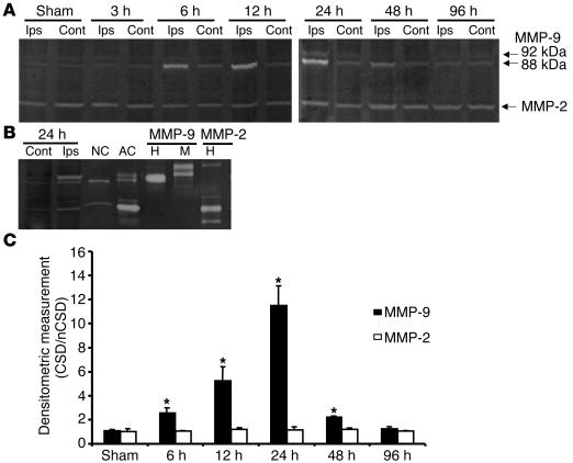
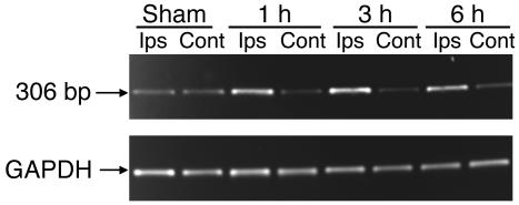
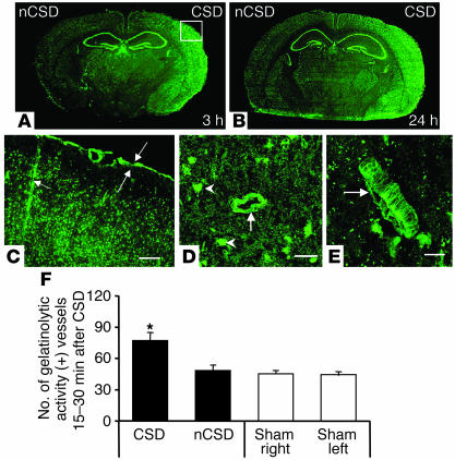
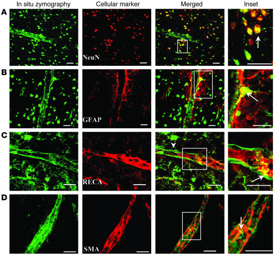
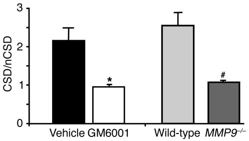
Cortical spreading depression (CSD) is a propagating wave of neuronal and glial depolarization and has been implicated in disorders of neurovascular regulation such as stroke, head trauma, and migraine. In this study, we found that CSD alters blood-brain barrier (BBB) permeability by activating brain MMPs. Beginning at 3-6 hours, MMP-9 levels increased within cortex ipsilateral to the CSD, reaching a maximum at 24 hours and persisting for at least 48 hours. Gelatinolytic activity was detected earliest within the matrix of cortical blood vessels and later within neurons and pia arachnoid (> or =3 hours), particularly within piriform cortex; this activity was suppressed by injection of the metalloprotease inhibitor GM6001 or in vitro by the addition of a zinc chelator (1,10-phenanthroline). At 3-24 hours, immunoreactive laminin, endothelial barrier antigen, and zona occludens-1 diminished in the ipsilateral cortex, suggesting that CSD altered proteins critical to the integrity of the BBB. At 3 hours after CSD, plasma protein leakage and brain edema developed contemporaneously. Albumin leakage was suppressed by the administration of GM6001. Protein leakage was not detected in MMP-9-null mice, implicating the MMP-9 isoform in barrier disruption. We conclude that intense neuronal and glial depolarization initiates a cascade that disrupts the BBB via an MMP-9-dependent mechanism.


Figures

Figure 1
Figure 1
CSD caused time-dependent increases in MMP-9 levels, as assessed by gelatin gel zymography. Unilateral CSD was elicited by pinprick in the frontal cortex as described (see Methods). (A) MMP-9 gelatinolytic activity was consistently higher on the CSD side from 6 to 48 hours and returned to basal levels by 96 hours. The molecular weights of both bands corresponded to those of rat MMP-9 (92 kDa and 88 kDa). (B) Comparison of MMP-9 responses induced by CSD versus MMP-9 in conditioned media from rat cortical neuron (NC) and astrocyte (AC) cell cultures incubated with 100 nM phorbol myristate acetate (PMA) for 24 hours. 92 kDa recombinant human MMP-9 (H) and 105 kDa recombinant mouse MMP-9 (M) standards were also loaded as molecular weight markers. MMP-9 bands induced by CSD at 24 hours matched those obtained from neuron and astrocyte cultures. Ips, ipsilateral (CSD); Cont, contralateral (non-CSD). (C) Densitometric measurements confirm the increase in MMP-9 induced by CSD. Constitutive MMP-2 expression and activity did not change after CSD and were visualized in sham as well. nCSD, non-CSD. *P < 0.05 compared with the contralateral side.
Figure 2
Figure 2
CSD upregulated MMP-9 mRNA in piriform cortex at 1, 3, and 6 hours. The housekeeping gene GAPDH is shown for comparison. MMP-9 mRNA was constitutively expressed in both brain cortices from control and contralateral to CSD.
Figure 3
Figure 3
The NMDA receptor blocker MK-801 blocked CSD and suppressed MMP-9 activity. (A) Topical MK-801 was administered 15 minutes before CSD. No change in MMP-2 expression was detected. These results suggest that MMP-9 activity is related to CSD but not to the needle stab (n = 3). (B) The nonselective metalloprotease inhibitor GM6001 was administered intraperitoneally 1 and 4 hours after CSD (65 mg/kg for each). A marked reduction in MMP-9 activity was found by gel zymography 12 hours later (two of three samples showed decreased or no MMP level). These results indicate that CSD increased MMP activity in vivo.
Figure 4
Figure 4
CSD increased in situ gelatinolytic activity in cortex ipsilateral to CSD. (A_E) Activity appeared as green fluorescent product and developed after incubation of coronal sections (10 ∝m in thickness) with the fluorogenic substrate DQ gelatin. Increased gelatinolytic activity was detected at 3 hours (A) and 24 hours (B). The image in C represents a higher magnification of the boxed area in A (scale bar: 100 ∝m). C shows gelatinolytic activity in penetrating blood vessels (single arrow) and in pia arachnoid (opposing arrows). (D and E) Activity is visualized in a blood vessel (arrow) cut in cross-section (D) and in a vessel (arrow) showing corrugations (E). Scale bars: 20 ∝m. Gelatinolytic activity was inhibited by preincubation with 1,10-phenanthroline (a nonselective zinc chelator) (data not shown). (F) In situ gelatinolytic activity was observed around blood vessels as early as 30 minutes to 1 hour after a single CSD, and the number of labeled vessels was significantly higher on the CSD side than on the non-CSD side or in sham-treated animals (*P < 0.05 compared with non-CSD side or sham cortices for right and left hemispheres).
Figure 5
Figure 5
CSD increased gelatinolytic activity within the vascular matrix. (A_D) Distribution of DQ gelatin_cleaving activity (in situ zymography) was compared on single sections, with the locations of cells identified by staining with specific cell markers: astrocytes, GFAP; neurons, NeuN; endothelial cells, RECA; smooth muscle cells, SMA. Cell markers were visualized by Cy-3 conjugated secondary antibody and were stained red in color. Boxed areas from merged confocal images (Merged column) are shown at higher magnification in the far right column (Inset). Gelatin cleaving activity was prominent in the walls of blood vessels and within surrounding neurons. Cleaving activity was rarely found colocalized with GFAP-positive cells. The figure shows one example. Infrequent activity was detected in endothelial cells and smooth muscle cells but predominantly within the vascular matrix. Sections were obtained 12 hours after CSD. Arrows point to areas showing colocalization. Scale bars: 20 ∝m.
Figure 6
Figure 6
CSD altered the immunostaining of laminin, ZO-1, and EBA within the blood vessel wall. (A_C) Laminin (approximate molecular weight, 200 kDa), a basement membrane protein, was reduced ipsilateral to CSD on immunoblots (at 12 hours) compared with that of the sham group and compared with that of the contralateral side. Similarly, CSD decreased the immunostaining of laminin in blood vessels (C), indicating that CSD alters the antigenicity of an integral basement membrane protein (scale bars: 100 ∝m). (B) These images show colocalization of the tight junction protein ZO-1 with RECA and show that CSD decreases the extent of this colocalization (colocalization coefficient, 0.21 ± 0.03 for CSD versus 0.55 ± 0.05 for control; P < 0.05). Compared with the image of the opposite side (nCSD), the merged image after CSD shows large segments of blood vessels with green fluorescence only (arrows; scale bars: 20 ∝m). (C) EBA immunostaining was also reduced in cortical blood vessels after CSD. This decrease often indicates impaired BBB function (scale bars: 100 ∝m).
Figure 7
Figure 7
CSD causes Evans blue leakage and edema within cerebral cortex. (A) Evans blue was injected 15 minutes prior to the onset of CSD. The vital dye (Evans blue) was extracted from cortical tissues at the indicated time points. There was a statistically significant difference (*, see below) between leakage into the CSD cortex and that of the sham group or the contralateral side. (B) Plasma proteins leak from cortical blood vessels after CSD. The number of vessels (>10 ∝m in diameter) in which leakage was detected on both CSD and non-CSD (contralateral) cortex are shown for six animals (P < 0.05). Confocal images show one vessel leaking Evans blue on the CSD side and a non-leaking vessel on the non-CSD side (scale bars: 100 ∝m). (C) Evans blue leakage was long lasting. When injected at 21 hours after CSD, vital dye was detected in CSD cortex 3 hours later and it was higher than that of sham cortex and the non-CSD side (n = 5; P < 0.05). (D) CSD caused a time-dependent increase in edema, as expressed by percentage water content (wet-dry/wet) brain weights. At 3, 6, and 24 hours, edema measured in piriform cortex increased on the CSD side compared with that of the non-CSD side and sham group. Asterisk denotes significant difference compared with contralateral cortex or to the sham group in A_D; P < 0.05.
Figure 8
Figure 8
CSD-induced plasma protein leakage was suppressed after MMP inhibition or after CSD in mice lacking MMP-9 gene expression. Evans blue leakage is blocked by the nonselective metalloprotease inhibitor GM6001 (65 mg/kg intraperitoneally, given 1 hour before and 4 hours after CSD) when examined at 12 hours, compared with that of vehicle-treated animals (n = 5 per group; *P < 0.05 vs. vehicle). A decrease in leakage 6 hours after CSD was detected in mice lacking MMP-9 gene expression compared with that of the same age-matched mice of similar background (CD-1) (n = 5 per group; #P < 0.05 vs. wild-type strain), although CSD itself did not differ between strains. Data are expressed as a ratio of Evans blue leakage in homogenates from both sides.

References

    1. Sugaya E, Takato M, Noda Y. Neuronal and glial activity during spreading depression in cerebral cortex of cat. J. Neurophysiol. 1975;38:822–841. - PubMed
    1. Kager H, Wadman WJ, Somjen GG. Simulated seizures and spreading depression in a neuron model incorporating interstitial space and ion concentrations. J. Neurophysiol. 2000;84:495–512. - PubMed
    1. Leao AAP. Pial circulation and spreading depression activity in cerebral cortex. J. Neurophysiol. 1944;7:391–396.
    1. Pantoni L, Lamassa M, Inzitari D. Transient global amnesia: a review emphasizing pathogenic aspects. Acta Neurol. Scand. 2000;102:275–283. - PubMed
    1. Martins-Ferreira H, Nedergaard M, Nicholson C. Perspectives on spreading depression. Brain Res. Brain Res. Rev. 2000;32:215–234. - PubMed

Publication types