

Topology of the substrate-binding site of a Lys49-phospholipase A2 influences Ca2+-independent membrane-damaging activity
- PMID: 15147240
- PMCID: PMC1133930
- DOI: 10.1042/BJ20031946
Topology of the substrate-binding site of a Lys49-phospholipase A2 influences Ca2+-independent membrane-damaging activity
Abstract
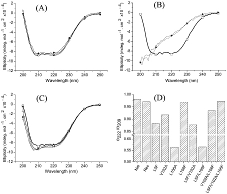
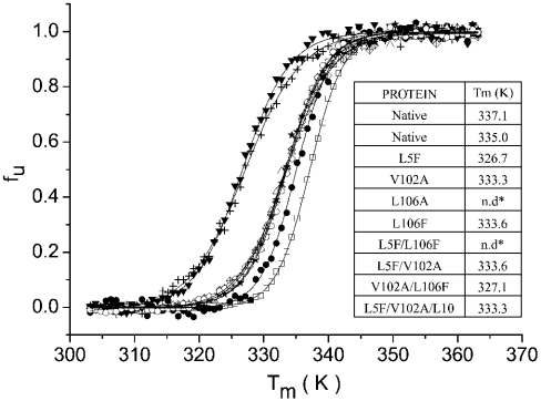
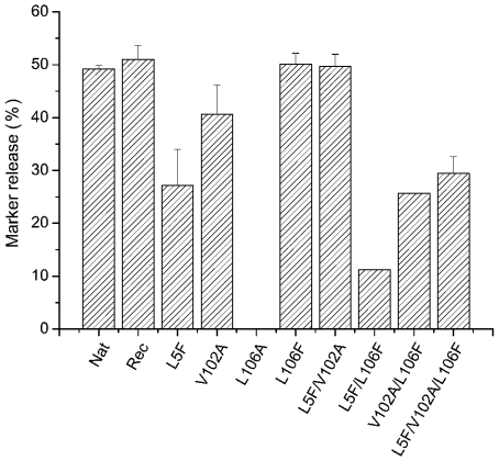
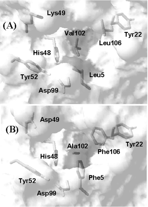
BthTx-I (bothropstoxin-I) is a myotoxic Lys49-PLA2 (phospholipase A2 with Lys49) isolated from Bothrops jararacussu venom, which damages liposome membranes by a Ca2+-independent mechanism. The highly conserved Phe5/Ala102/Phe106 motif in the hydrophobic substrate-binding site of the Asp49-PLA2s is substituted by Leu5/Val102/Leu106 in the Lys49-PLA2s. The Leu5/Val102/Leu106 triad in BthTx-I was sequentially mutated via all single- and double-mutant combinations to the Phe5/Ala102/Phe106 mutant. All mutants were expressed as inclusion bodies in Escherichia coli, and the thermal stability (Tm), together with the myotoxic and Ca2+-independent membrane-damaging activities of the recombinant proteins, were evaluated. The far-UV CD profiles of the native, wild-type recombinant and the L106F (Leu106-->Phe) and L5F/F102A/L106F mutant proteins were identical. The L5F, V102A, L5F/V102A and V102A/L106F mutants showed distorted far-UV CD profiles; however, only the L5F and L5F/V102A mutants showed significant decreases in Tm. Alterations in the far-UV CD spectra correlated with decreased myotoxicity and protein-induced release of a liposome-entrapped marker. However, the V102A/L106F and L5F/V102A/L106F mutants, which presented high myotoxic activities, showed significantly reduced membrane-damaging activity. This demonstrates that the topology of the substrate-binding region of BthTx-I has a direct effect on the Ca2+-independent membrane damage, and implies that substrate binding retains an important role in this process.
Figures

Similar articles
-
Active-site mutagenesis of a Lys49-phospholipase A2: biological and membrane-disrupting activities in the absence of catalysis.Biochem J. 2002 Feb 15;362(Pt 1):89-96. doi: 10.1042/0264-6021:3620089. Biochem J. 2002. PMID: 11829743 Free PMC article.
-
Refolding and purification of Bothropstoxin-I, a Lys49-phospholipase A2 homologue, expressed as inclusion bodies in Escherichia coli.Protein Expr Purif. 2001 Feb;21(1):134-40. doi: 10.1006/prep.2000.1353. Protein Expr Purif. 2001. PMID: 11162398
-
Distinct sites for myotoxic and membrane-damaging activities in the C-terminal region of a Lys49-phospholipase A2.Biochem J. 2002 Sep 15;366(Pt 3):971-6. doi: 10.1042/BJ20020092. Biochem J. 2002. PMID: 12079495 Free PMC article.
-
An overview of lysine-49 phospholipase A2 myotoxins from crotalid snake venoms and their structural determinants of myotoxic action.Toxicon. 2003 Dec 15;42(8):885-901. doi: 10.1016/j.toxicon.2003.11.008. Toxicon. 2003. PMID: 15019489 Review.
-
Mapping structural determinants of biological activities in snake venom phospholipases A2 by sequence analysis and site directed mutagenesis.Toxicon. 2003 Dec 15;42(8):869-83. doi: 10.1016/j.toxicon.2003.11.027. Toxicon. 2003. PMID: 15019488 Review.
Cited by
-
Neurotoxicity and other pharmacological activities of the snake venom phospholipase A2 OS2: the N-terminal region is more important than enzymatic activity.Biochemistry. 2006 May 9;45(18):5800-16. doi: 10.1021/bi060217r. Biochemistry. 2006. PMID: 16669624 Free PMC article.
References
-
- van Deenan L. L. M., de Haas G. H. The substrate specificity of phospholipase A2. Biochim. Biophys. Acta. 1963;70:538–553. - PubMed
-
- Six D. A., Dennis E. A. The expanding superfamily of phospholipase A(2) enzymes: classification and characterization. Biochim. Biophys. Acta. 2000;1488:1–19. - PubMed
-
- Verheij H. M., Volwerk J. J., Jansen E. H. J. M., Puyk W. C., Dijkstra B. W., Drenth J., de Haas G. H. Methylation of histidine-48 in pancreatic phospholipase A2. Role of histidine and calcium ion in the catalytic mechanism. Biochemistry. 1980;19:743–750. - PubMed
-
- Gutierrez J. M., Lomonte B. Phospholipase A2 myotoxins from Bothrops snake venoms. Toxicon. 1995;33:1405–1424. - PubMed
Publication types
MeSH terms
Substances
LinkOut - more resources
Full Text Sources
Miscellaneous