

Chondroitin sulphate structure affects its immunological activities on murine splenocytes sensitized with ovalbumin
- PMID: 15147241
- PMCID: PMC1133940
- DOI: 10.1042/BJ20031851
Chondroitin sulphate structure affects its immunological activities on murine splenocytes sensitized with ovalbumin
Abstract
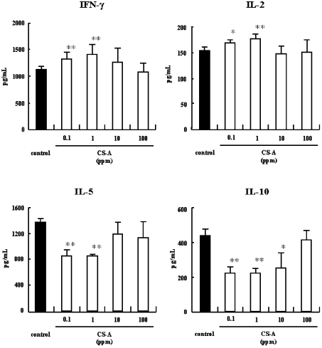
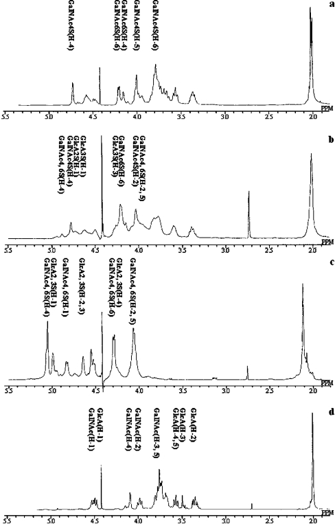
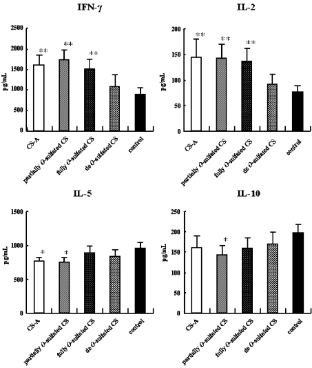
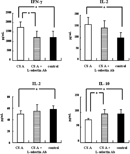
Chondroitin sulphate (CS) is a glycosaminoglycan widely distributed in animal tissues, which has anti-inflammatory and chondroprotective properties. We reported previously that chondroitin 4-sulphate (CS-A) up-regulates the antigen-specific Th1 immune response of murine splenocytes sensitized with ovalbumin in vitro, and that CS suppresses the antigen-specific IgE responses. We now demonstrate that a specific sulphation pattern of the CS polysaccharide is required for the Th1-promoted activity, as other polysaccharides such as dextran and dextran sulphate do not significantly induce this activity. While the presence of some O-sulpho groups appear to be essential for activity, CS-A, and synthetically prepared, partially O-sulphonated CS, induce higher Th1-promoted activity than synthetically prepared, fully O-sulphonated CS. CS-A induces an activity greater than chondroitin sulphate B (CS-B) or chondroitin 6-sulphate (CS-C). In addition, chondroitin sulphate E (CS-E) induces greater activity than CS-A or CS-D. These results suggest that the GlcA(beta1-3)GalNAc(4,6-O-disulpho) sequence in CS-E is important for Th1-promoted activity. Furthermore, rat anti-mouse CD62L antibody, an antibody to L-selectin, inhibits the Th1-promoting activity of CS. These results suggest that the Th1-promoted activity could be associated with L-selectin on lymphocytes. These findings describe a new mechanism for the anti-inflammatory and chondroprotective properties of CS that may be useful in designing new therapeutic applications for CS used in the treatment of immediate-type hypersensitivity.
Figures


Similar articles
-
Chondroitin sulfate intake inhibits the IgE-mediated allergic response by down-regulating Th2 responses in mice.J Biol Chem. 2006 Jul 21;281(29):19872-80. doi: 10.1074/jbc.M509058200. Epub 2006 Apr 19. J Biol Chem. 2006. PMID: 16624819 Free PMC article.
-
Effect of chondroitin sulfate on murine splenocytes sensitized with ovalbumin.Immunol Lett. 2002 Dec 3;84(3):211-6. doi: 10.1016/s0165-2478(02)00181-5. Immunol Lett. 2002. PMID: 12413739
-
Mechanistic insights into cellular immunity of chondroitin sulfate A and its zwitterionic N-deacetylated derivatives.Carbohydr Polym. 2015 Jun 5;123:331-8. doi: 10.1016/j.carbpol.2015.01.059. Epub 2015 Feb 3. Carbohydr Polym. 2015. PMID: 25843866
-
Biosynthesis and function of chondroitin sulfate.Biochim Biophys Acta. 2013 Oct;1830(10):4719-33. doi: 10.1016/j.bbagen.2013.06.006. Epub 2013 Jun 14. Biochim Biophys Acta. 2013. PMID: 23774590 Review.
-
Immunomodulatory and anti-inflammatory effects of chondroitin sulphate.J Cell Mol Med. 2009 Aug;13(8A):1451-63. doi: 10.1111/j.1582-4934.2009.00826.x. Epub 2009 Jun 11. J Cell Mol Med. 2009. PMID: 19522843 Free PMC article. Review.
Cited by
-
Chondroitin sulfate intake inhibits the IgE-mediated allergic response by down-regulating Th2 responses in mice.J Biol Chem. 2006 Jul 21;281(29):19872-80. doi: 10.1074/jbc.M509058200. Epub 2006 Apr 19. J Biol Chem. 2006. PMID: 16624819 Free PMC article.
-
Regulation of Macrophage and Dendritic Cell Function by Chondroitin Sulfate in Innate to Antigen-Specific Adaptive Immunity.Front Immunol. 2020 Mar 3;11:232. doi: 10.3389/fimmu.2020.00232. eCollection 2020. Front Immunol. 2020. PMID: 32194548 Free PMC article. Review.
-
Development of Polyelectrolyte Complexes for the Delivery of Peptide-Based Subunit Vaccines against Group A Streptococcus.Nanomaterials (Basel). 2020 Apr 26;10(5):823. doi: 10.3390/nano10050823. Nanomaterials (Basel). 2020. PMID: 32357402 Free PMC article.
-
Immune modulation by chondroitin sulfate and its degraded disaccharide product in the development of an experimental model of multiple sclerosis.J Neuroimmunol. 2010 Jun;223(1-2):55-64. doi: 10.1016/j.jneuroim.2010.04.002. J Neuroimmunol. 2010. PMID: 20434781 Free PMC article.
-
Immunological activity of chondroitin sulfate.Adv Pharmacol. 2006;53:403-15. doi: 10.1016/S1054-3589(05)53019-9. Adv Pharmacol. 2006. PMID: 17239777 Free PMC article. Review. No abstract available.
References
-
- Roden L. New York: Plenum Press; 1980. The biochemistry of glycoproteins and proteoglycans; pp. 267–371.
-
- Kimata K., Okayama M., Oohira A., Suzuki S. Cytodifferentiation and proteoglycan biosynthesis. Mol. Cell. Biochem. 1973;1:211–228. - PubMed
-
- Toida T., Toyoda H., Imanari T. High-resolution proton nuclear magnetic resonance studies on chondroitin sulphates. Anal. Sci. 1993;9:53–58.
-
- Scott J. E. Structure and function in extracellular matrices depend on interactions between anionic glycosaminoglycans. Pathol. Biol. (Paris) 2001;49:284–289. - PubMed
Publication types
MeSH terms
Substances
Grants and funding
LinkOut - more resources
Full Text Sources