Comparative Study
. 2004 May;19(10):2741-52.
doi: 10.1111/j.0953-816X.2004.03366.x.

Effects of hypocretin2-saporin and antidopamine-beta-hydroxylase-saporin neurotoxic lesions of the dorsolateral pons on sleep and muscle tone

Affiliations
Comparative Study

Effects of hypocretin2-saporin and antidopamine-beta-hydroxylase-saporin neurotoxic lesions of the dorsolateral pons on sleep and muscle tone

Carlos Blanco-Centurion et al. Eur J Neurosci. 2004 May.

Abstract

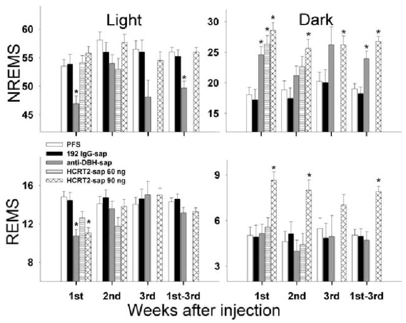
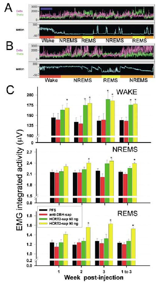
The hypocretin neurons have been implicated in regulating sleep-wake states as they are lost in patients with the sleep disorder narcolepsy. Hypocretin (HCRT) neurons are located only in the perifornical region of the posterior hypothalamus and heavily innervate pontine brainstem neurons, such as the locus coeruleus (LC), which have traditionally been implicated in promoting arousal. It is not known how the hypocretin innervation of the pons regulates sleep-wake states as pontine lesions have never been shown to increase sleep. It is likely that in previous studies specific neurons were not lesioned. Therefore, in this study, we applied saporin-based neurotoxins to the dorsolateral pons and monitored sleep in rats. Anti-dopamine-beta-hydroxylase-saporin killed the LC neurons but sleep was affected only during a two hour light-dark transition period. Application of hypocretin2-saporin killed fewer LC neurons relative to other adjacent neurons. This occurred because the LC neurons possess the hypocretin receptor 1 but the ligand hypocretin 2 binds to this receptor with less affinity relative to the hypocretin receptor 2. The hypocretin2-saporin lesioned rats compared to controls had increased sleep during the dark period and displayed increased limb movements during REM sleep. None of the lesioned rats had sleep onset REM sleep periods or cataplexy. We conclude that the hypocretin innervation to the pons functions to awaken the animal when the lights turn off (via its innervation of the LC), sustains arousal and represses sleep during the rest of the night (via a wider innervation of other pontine neurons), and modulates muscle tone.


Figures

Fig. 1
Fig. 1
Effects of anti-DBH-sap on noradrenergic neurons in the LC. Photo A depicts a coronal section of the pons containing the LC in a rat administered with PFS. Panel D is an enlargement of the LC nucleus from the same rat shown in A. Panels B and C represent two examples of LC lesions following a unilateral (panel B) or bilateral microinjection of anti-DBH-sap in the LC. The tissue was reacted for visualization of DBH immunoreactivity and NADPH-diaphorase activity (to identify cholinergic neurons). Cells stained in black were positive for dopamine-β-hydroxylase whereas blue stained cells (evident in photos D and E and highlighted by arrows) represent NADPH-diaphorase activity. The tissue was also counterstained with neutral red (red cells). Anti-DBH-sap lesioned the noradrenergic LC neurons but not the adjacent cholinergic neurons (photo E). Scale bar in B applies to A and C and scale bar in E applies to D.
Fig. 2
Fig. 2
Effects of saporin-conjugated neurotoxins on number of noradrenergic LC neurons (DBH-ir+) and cholinergic (identified as NADPH+) neurons in the lateral dorsal tegmental nucleus. ***P< 0.001 vs. PFS, *P< 0.015 vs. PFS.
Fig. 3
Fig. 3
Effects of anti-DBH-sap on the expression of two proteins normally present in LC neurons. Photos A and B identify LC neurons labelled with tyrosine hydroxylase. Photo A identifies the LC in the side contralateral to the anti-DBH-sap injection while photo B is from the side given anti-DBH-sap in the LC. Photo C identifies NeuN labelled neurons in the LC and adjacent dorsolateral pontine tegmentum. In photo C, no NeuN labelled neurons are evident in the LC receiving anti-DBH-sap relative to the contralateral side (LC is marked by an arrow in photo C) indicating that the neurotoxin killed LC neurons rather than only inhibiting the activity of the enzyme DBH present in LC neurons.
Fig. 4
Fig. 4
Effects of anti-DBH-sap lesions of the LC on percentage wake, NREMS and REMS over a 3-week period. The data are double plotted, with the dark bar representing the dark period. The primary effect at 3 weeks after anti-DBH-sap injection was during the light–dark transition period. The lesioned animals were awake more (and had less NREMS) during the two-hour period after the lights turned on, and they were awake less (and had more NREMS) when the lights turned off. Asterisks denote P < 0.05 anova. PFS n = 6, anti-DBH-sap n = 5.
Fig. 5
Fig. 5
Effects of saporin-conjugated neurotoxins on sleep-wake states (mean percentage ± SEM) during the 12-h light and dark periods. There was no data from HCRT2-sap 60 ng during the third week as the sleep recording electrodes became disconnected from many of the rats in this group. 1st–3rd represents the average of the 3-week period. Asterisk denotes P < 0.001 vs. PFS treated rats.
Fig. 6
Fig. 6
Effects of HCRT2-sap on noradrenergic LC and cholinergic LDT-PPT neurons. HCRT2-sap killed more cholinergic neurons relative to noradrenergic LC neurons. Photo A depicts a coronal section of the dorsolateral pons labelled to identify DBH-ir neurons in the LC. The tissue section is from a rat that received HCRT2-sap (90ng/μL) 3 weeks earlier, but the LC is still visible. Panels B and C depict NADPH positive cells in the LDT (B) as well as PPT region (C) of a rat with a PFS injection in the LC. Photos D and E are showing how after HCRT2-sap (90 ng/μL) injection there is a significant loss of LDT cholinergic neurons (D) but not of PPT cholinergic neurons (E). D and E are tissue sections taken from the same rat depicted in A. Cholinergic neurons were identified by NADPH-staining. LDT, lateral dorsal tegmental nucleus; scp, superior cerebellar peduncle; PPT, pedunculopontine tegmental nucleus.
Fig. 7
Fig. 7
Effects of HCRT2-sap on NeuN labelled neurons. Photomicrographs are from rats given PFS (panel A) or HCRT2-sap 90 ng/μL (panel B and C) aimed at the locus coeruleus (LC). HCRT2-sap produced a loss of neurons within subcoeruleus alpha (SCα), the medial parabrachial nucleus (MPB), the medial pontine reticular formation, the superior cerebellar peduncle (scp), lateral parabrachial nucleus (LPB) and laterodorsal tegmental nucleus (LDT). When adjacent sections were stained to identify noradrenergic and cholinergic neurons there was a clear loss of cholinergic neurons but not of the LC neurons (see Figs 2 and 6). Panel C is a higher magnification of the lesioned area delineated as a rectangle in B and there are visible neurons within the lesion zone, indicating that even within the lesion area some neurons are spared, perhaps because they did not possess the appropriate HCRT receptor. DMTg, dorsomedial tegmental area; DTg, dorsal tegmental nucleus.
Fig. 8
Fig. 8
Extent of the lesion in the dorsolateral pontine tegmentum produced by the two concentrations of HCRT2-sap. Substantial neuronal loss (shown by loss of NeuN-ir) as well as gliosis response (shown by neutral red) were used to outline lesion areas one month after the injection. Figures depict the rostral (left column), central (middle column) and caudal extents (right column) of the lesion in every rat that received HCRT2-sap (90 ng, n = 6; 60 ng, n = 5). A number located at top left of every series identifies individual rats. Numbers at the bottom of every coronal section indicate the anterior–posterior planes from the interaural line and correspond to drawings from the Paxinos & Watson’s rat brain atlas, (Paxinos & Watson, 1986).
Fig. 9
Fig. 9
Effects of HCRT2-sap (90ng/μL) on percentage wake, NREMS and REM sleep. The data (means ± SEM) are double-plotted with the abscissa indicating time in 2-h blocks. Open and closed horizontal bars indicate the light and dark periods. *P < 0.05 anova with Tukey’s test. PFS n = 6, HCRT2-sap n = 6.
Fig. 10
Fig. 10
Effects of HCRT2-sap on integrated muscle tone during REM sleep. Panels A and B depict 2 min segments of a sleep-wake recording in rats given PFS (panel A) or HCRT2-sap (panel B) to the dorsolateral pons. Each panel (A and B) consists of a recording of power of the EEG in the delta (0.3–4 Hz, magenta trace) and theta bands (4–12 Hz, green trace), and integrated activity of the nuchal muscles (Motion, light blue). The sleep-wake state determination, based on the relationship of the EEG (not shown here), power and EMG activity, is indicated at the bottom of each panel. Rats given HCRT2-sap displayed increased muscle tone during REM sleep (panel B). It was not unusual to witness rats displaying tread behaviour while in REM sleep, a phenomenon akin to REM sleep behaviour disorder. Panel C depicts the effects of HCRT2-sap or anti-DBH-sap on integrated muscle tone during specific behavioural states over the 3-week recording period. EMG activity was recorded from flexible wires inserted in the nuchal muscles, and the integrated EMG activity (integrated every second and averaged over 12 s) is represented in the ordinate scale. EMG data are means ± SEM. PFS n = 6, anti-DBH-sap n = 5, HCRT2-sap 60 ng/μL n = 5, HCRT2-sap 90 ng/μL n = 6. *P< 0.05 vs. PFS. Dark blue bar located on the left corner at the top panel indicates 2 min.

References

    1. Aston-Jones G, Chen S, Zhu Y, Oshinsky ML. A neural circuit for circadian regulation of arousal. Nature Neurosci. 2001;4:732–738. - PubMed
    1. Blessing WW, Lappi DA, Wiley RG. Destruction of locus coeruleus neuronal perikarya after injection of anti-dopamine-B-hydroxylase immunotoxin into the olfactory bulb of the rat. Neurosci Lett. 1998;243:85–88. - PubMed
    1. Bourgin P, Escourrou P, Gaultier C, Adrien J. Induction of rapid eye movement sleep by carbachol infusion into the pontine reticular formation in the rat. Neuroreport. 1995;6:532–536. - PubMed
    1. Bourgin P, Huitron-Resendiz S, Spier AD, Fabre V, Morte B, Criado JR, Sutcliffe JG, Henriksen SJ, De Lecea L. Hypocretin-1 modulates rapid eye movement sleep through activation of locus coeruleus neurons. J Neurosci. 2000;20:7760–7765. - PMC - PubMed
    1. Bremer F. Isolated brain and physiology of sleep. CR Soc Biol. 1935;118:1235–1241.

Publication types

MeSH terms