Identification of systemically expanded activated T cell clones in MRL/lpr and NZB/W F1 lupus model mice
- PMID: 15147346
- PMCID: PMC1809066
- DOI: 10.1111/j.1365-2249.2004.02473.x
Identification of systemically expanded activated T cell clones in MRL/lpr and NZB/W F1 lupus model mice
Abstract
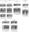
CD4(+) T lymphocytes play an important role in the pathogenesis of systemic lupus erythematosus (SLE). To characterize the clonal expansion of CD4(+) T cells in murine lupus models, we analysed the T cell clonality in various organs of young and nephritic MRL/lpr and NZB/W F1 mice using reverse transcription-polymerase chain reaction (RT-PCR) and subsequent single-strand conformation polymorphism (SSCP) analysis. We demonstrated that some identical T cell clonotypes expanded and accumulated in different organs (the bilateral kidneys, brain, lung and intestine) in nephritic diseased mice, and that a number of these identical clonotypes were CD4(+) T cells. In contrast, young mice exhibited little accumulation of common clones in different organs. The T cell receptor (TCR) V beta usage of these identical clonotypes was limited to V beta 2, 6, 8.1, 10, 16 and 18 in MRL/lpr mice and to V beta 6 and 7 in NZB/W F1 mice. Furthermore, some conserved amino acid motifs such as I, D or E and G were observed in CDR3 loops of TCR beta chains from these identical CD4(+) clonotypes. The existence of systemically expanding CD4(+) T cell clones in the central nervous system (CNS) suggests the involvement of the systemic autoimmunity in CNS lesions of lupus. FACS-sorted CD4(+)CD69(+) cells from the kidney displayed expanded clonotypes identical to those obtained from the whole kidney and other organs from the same individual. These findings suggest that activated and clonally expanded CD4(+) T cells accumulate in different tissues of nephritic lupus mice, and these clonotypes might recognize restricted T cell epitopes on autoantigens involved in specific immune responses of SLE, thus playing a pathogenic role in these lupus mice.
Figures


Similar articles
-
Clonal expansion of CD4+ TCRbetabeta+ T cells in TCR alpha-chain- deficient mice by gut-derived antigens.J Immunol. 1999 Feb 1;162(3):1843-50. J Immunol. 1999. PMID: 9973450
-
T cell receptor V beta genes expressed by IgG anti-DNA autoantibody-inducing T cells in lupus nephritis: forbidden receptors and double-negative T cells.Eur J Immunol. 1990 Jul;20(7):1435-43. doi: 10.1002/eji.1830200705. Eur J Immunol. 1990. PMID: 2143726
-
A subset of CD4+ T cells expressing early activation antigen CD69 in murine lupus: possible abnormal regulatory role for cytokine imbalance.J Immunol. 1998 Aug 1;161(3):1267-73. J Immunol. 1998. PMID: 9686587
-
Anti-nucleosome antibodies and T-cell response in systemic lupus erythematosus.Ann Med Interne (Paris). 2002 Dec;153(8):513-9. Ann Med Interne (Paris). 2002. PMID: 12610425 Review.
-
Altered expression of the T cell receptor-CD3 complex in systemic lupus erythematosus.Int Rev Immunol. 2004 May-Aug;23(3-4):273-91. doi: 10.1080/08830180490452594. Int Rev Immunol. 2004. PMID: 15204089 Review.
Cited by
-
Composition and variation analysis of TCR β-chain CDR3 repertoire in the thymus and spleen of MRL/lpr mouse at different ages.Immunogenetics. 2015 Jan;67(1):25-37. doi: 10.1007/s00251-014-0809-y. Epub 2014 Oct 22. Immunogenetics. 2015. PMID: 25335895
-
Therapy for pneumonitis and sialadenitis by accumulation of CCR2-expressing CD4+CD25+ regulatory T cells in MRL/lpr mice.Arthritis Res Ther. 2007;9(1):R15. doi: 10.1186/ar2122. Arthritis Res Ther. 2007. PMID: 17284325 Free PMC article.
-
Prenatal Administration of Betamethasone Causes Changes in the T Cell Receptor Repertoire Influencing Development of Autoimmunity.Front Immunol. 2017 Nov 13;8:1505. doi: 10.3389/fimmu.2017.01505. eCollection 2017. Front Immunol. 2017. PMID: 29181000 Free PMC article.
-
The role of dendritic cells in the mechanism of action of a peptide that ameliorates lupus in murine models.Immunology. 2009 Sep;128(1 Suppl):e395-405. doi: 10.1111/j.1365-2567.2008.02988.x. Epub 2008 Nov 24. Immunology. 2009. PMID: 19040426 Free PMC article.
-
Longitudinal analysis of peripheral blood T cell receptor diversity in patients with systemic lupus erythematosus by next-generation sequencing.Arthritis Res Ther. 2015 May 23;17(1):132. doi: 10.1186/s13075-015-0655-9. Arthritis Res Ther. 2015. PMID: 26001779 Free PMC article.
References
-
- Rozzo SJ, Drake CG, Chiang BL, et al. Evidence for polyclonal T cell activation in murine models of systemic lupus erythematosus. J Immunol. 1994;153:1340–51. - PubMed
-
- Giese T, Davidson WF. Evidence for early onset, polyclonal activation of T cell subsets in mice homozygous for lpr. J Immunol. 1992;149:3097–106. - PubMed
-
- Koh DR, Ho A, Rahemtulla A, et al. Murine lupus in MRL/lpr mice lacking CD4 or CD8 T cells. Eur J Immunol. 1995;25:2558–62. - PubMed
-
- Jabs DA, Burek CL, Hu Q, et al. Anti-CD4 monoclonal antibody therapy suppresses autoimmune disease in MRL/Mp-lpr/lpr mice. Cell Immunol. 1992;141:496–507. - PubMed
Publication types
MeSH terms
Substances
LinkOut - more resources
Full Text Sources
Medical
Research Materials

