. 2004 Jun;136(3):472-82.
doi: 10.1111/j.1365-2249.2004.02472.x.

Recognition of nonpeptide antigens by human V gamma 9V delta 2 T cells requires contact with cells of human origin

Affiliations

Recognition of nonpeptide antigens by human V gamma 9V delta 2 T cells requires contact with cells of human origin

A E Green et al. Clin Exp Immunol. 2004 Jun.

Abstract

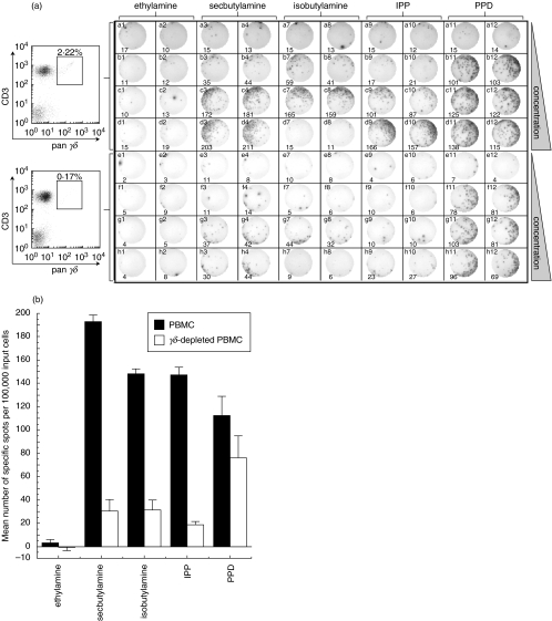
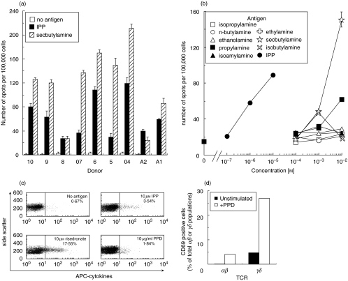
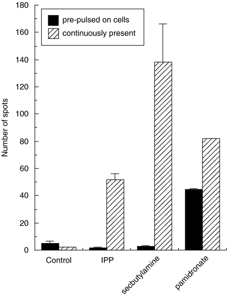
SUMMARY It is becoming apparent that gamma delta T cells form an important part of the adaptive immune response. However, the ligands recognized by gamma delta T cell receptors (TCRs) and the exact biological function of the cells that express this receptor remain unclear. Numerous studies have shown that the dominant human peripheral blood subset of gamma delta T cells, which express a V gamma 9V delta 2 TCR, can activate in response to low molecular weight nonpeptidic molecules. Some of these components have been purified from bacteria or parasites. We examined the activation of polyclonal gamma delta T cell lines, clones with V gamma 9V delta 2 and V gamma 9V delta 1 TCRs, and gamma delta T cells directly ex vivo in response to multiple phosphate, alkylamine and aminobisphosphonate (nBP) antigens and purified protein derivative from Mycobacterium tuberculosis (PPD). V gamma 9V delta 2 T cells were able to respond to multiple small organic molecules of highly variable structure whereas cells expressing a similar V gamma 9 chain paired with a V delta 1 chain failed to recognize these antigens. Thus, the TCR delta chain appears to make an important contribution to the recognition of these antigens. The kinetics of responses to alkylphosphate and alkylamine antigens differ from those of responses to the nBP pamidronate. These different classes of antigen are believed to have differed mechanisms of action. Such differences explain why nBPs can be pulsed onto antigen presenting cells (APCs) and still retain their ability to activate gamma delta T cells while alkylphosphate and alkylamine antigens cannot. We also demonstrate that a substantial proportion of the cells that produce IFN gamma directly ex vivo in response to PPD are gamma delta T cells and that gamma delta T cell activation requires contact with cells of human origin.


Figures

Fig. 1
Fig. 1
γδ T cells respond to alkylphosphate, alkylamine and PPD antigens directly ex vivo. (a) Demonstration of γδ T cell activation by direct ex vivo IFNγ ELISpot assay. The top half of the plate was set up with 100 000 PBMC/well and the bottom half with 100 000 γδ-depleted PBMC per well. FACS analysis with pan-γδ and anti-CD3 antibody (left) showed that γδ-depletion was >92% successful. Cells were activated with ethylamine in columns 1 & 2, secbutylamine in columns 3 & 4, isobutylamine in columns 7 & 8, IPP in columns 9 & 10 and PPD in columns 11 & 12. Two replicates were performed for each experimental condition. Rows a and e are control rows without added antigen. Alkylamine antigens were added at 1 mm in rows b & f, 10 mm in rows c & g and 50 mm in rows d & h. IPP was added at 10 µm in rows b & f, 100 µm in rows c & g and 1 mm in rows d & h. PPD was added at 5 µg/ml in rows b & f, 10 µg/ml in rows c & g and 20 µg/ml in rows d & h. The ELISpot photograph and number of spots (bottom of each panel) were generated mechanically using an ELISpot Reader System ELR02 (Autoimmun Diagnostika; Strassberg). (b) Number of specific spots (sample minus background) per 100 000 PBMC (▪) and γδ-depleted PBMC (□) induced by specified antigens. Bars represent spots induced by 50 mm ethylamine, 50 mm secbutylamine, 10 mm isobutylamine (50 mm isobutylamine was toxic to cells), 1 mm IPP and 20 µg/ml PPD. Error bars show the standard deviation from the mean of two replicate wells.
Fig. 2
Fig. 2
Further characterization of direct ex vivo responses to γδ T cell antigens. (a) IFNγ ELISpot reponses to 100 µm IPP and 10 mm secbutylamine in PBMC from 9 separate individuals. All individuals also responded to n-butylamine, propylamine and isobutylamine (data not shown) although the response to these antigens was generally lower than that with secbutylamine. Bars show the standard deviation from the mean of two replicate assays. (b) The effect of antigen concentration on the activation of γδ T cells directly ex vivo. The number of spots per 100 000 input PBMC is shown. PBMC from other individuals gave similar results (data not shown). Isobutylamine is toxic at high concentration [32]. Ethylamine, isopropylamine, ethanolamine, and isoamylamine, previously reported to be antigenic [32], did not induce spots. Error bars show standard deviation from the mean of two replicate assays. The errors were generally smaller than the plot symbols. (c) 106 fresh PBMC from a BCG vaccinated healthy donor were exposed to indicated antigens for 6 h and then stained with anti-Vγ9 antibody and for intracellular cytokines (IFNγ, TNFα and IL2) as described in the Materials and Methods. Plots show side scatter vs. staining with APC-cytokine antibodies for the Vγ9 positive population and indicate the fraction of cytokine positive cells. (d) PPD induced CD69 up-regulation in almost 30% of human peripheral blood γδ T cells. PBMCs were washed in RPMI and resuspended in RPMI + 10% FCS at 106 cells/ml and incubated ± 10 µg/ml PPD at 37°C for 12 h. Cells were stained with FITC-conjugated anti-CD69 antibody, PE-conjugated anti-Vγ9 antibody and PerCP-conjugated anti-CD3 antibody prior to FACS analysis.
Fig. 3
Fig. 3
The γ and δ chain sequences of γδ T cell clones. The TCR sequences for the Vγ9Vδ1 (top box) and Vγ9Vδ2 (bottom box) T cell clones (IMGT nomenclature [36] is used throughout this work). The γ and δ chain sequences for the Vγ9Vδ1 clone have the accession numbers AJ583014 and AJ583015, respectively, at the EMBL Nucleotide Sequence Database. The accession numbers AJ583012 and AJ583013 have been assigned to the γ and δ chains for the Vγ9Vδ2 clone. The CDR1, 2 and 3 sequences are indicated by shaded boxes. The differences in the Vγ9 CDR3 region between the Vδ1 and Vδ2 clones are underlined. Both Vγ9 chains use the JγP joining region which contains two germline encoded lysine residues (shown in bold text within the CDR3 region) that have been shown to be essential for recognition of alkylphosphate and alkylamine ligands [46].
Fig. 4
Fig. 4
Vγ9Vδ1 and Vγ9Vδ2 T cells respond to different antigens. (a) Both the Vγ9Vδ1 (upper panels) and Vγ9Vδ2 (lower panels) T cell clones were shown to express high levels of TCR by FACS. Clones were stained with PE-conjugated anti-human Vγ9 mAb clone B3·1 (all panels), FITC-conjugated anti-human Vδ1 mAb clone TS8·2 (left panels) and FITC-conjugated anti-human Vδ2 mAb clone B6·1 (right panels). (b) Both clones can be activated through their TCR by anti-CD3 antibody. Clones were stimulated with anti-CD3 antibody (10 µg/ml) for 3 min, washed once with PBS, and lysed, before separation by SDS-PAGE alongside unstimulated controls. Proteins bearing phosphorylated tyrosine residues were revealed by immunoblotting with anti-phosphotyrosine antibody clone 4G10 as described [38]. The lysates of 106 cells per lane were loaded as follows: Vγ9Vδ1 T lymphocytes before (lane 1) and after (lane 2) stimulation; Vγ9Vδ2 T lymphocytes before (lane 3) and after (lane 4) stimulation. (c) Vγ9Vδ2, but not Vγ9Vδ1 T cells, are able to recognize alkylphosphate, alkylamine and nBP antigens. MIP1β release from 5 × 10⁴ clonal T cells as described in the Materials and Methods section is shown in response to 1·5 µg/ml anti-CD3 antibody clone UCHT-1, 10 µm IPP, 10 mm secbutylamine and 1 µm risedronate. (d) IFNγ ELISpot assay with a titration of potential antigens. 1000 Vγ9Vδ2 T cells were used with 25 000 EBV transformed B cells (Sparky line) per well as antigen presenting cells. The Vγ9Vδ1 clone failed to activate in response to any of these antigens (data not shown).
Fig. 5
Fig. 5
Alkylamine and alkylphosphate antigens exhibit different pharmacokinetics than nBP antigens. Mean number of spots induced by IPP and secbutylamine when 1000 cells from the Vγ9Vδ2 T cell clone Bob were incubated with 25 000 BCL line Sparky in the continuous presence (striped bars) of 100 µm IPP, 10 mm secbutylamine and 1 mm pamidronate during a 12 h IFNγ ELISpot. The filled bars are from a parallel assay where the BCL were incubated for 12 h in R10 containing the above concentrations of antigens and then washed once and resuspended in fresh media without antigen prior to the experiment. The ‘control’ targets were not pulsed with antigen. Bars show the standard deviation from the mean of two replicate assays. Only pamidronate could be ‘pulsed’ onto cells. Similar results were obtained with γδ T cell lines from three separate individuals (data not shown).
Fig. 6
Fig. 6
Recognition of alkylamine and alkylphosphate antigens by Vγ9Vδ2 T cells requires contact with cells of human origin. Mean number of spots in IFNγ ELISpot assay with and without antigen presenting cells. 1000 γδ T cells/well were plated with 25 000 EBV-transformed human B cells, Spinner Hela cells, rat myeloma Y3.AG.1.2.3 or mouse myeloma P3/NS1/1-Ag4·1. Only human cells were able to act as antigen presenting cells in this assay. The use of 25 000 γδ cells allowed cell-cell contact and confirms that these cells have some ability to present alkylphosphate and alkylamine antigens to each other. Secbutylamine and isobutylamine were added to assays at 10 mm; IPP was added at 100 µm. Assays were performed over 12 h as described in the Materials and Methods. Standard deviation from the mean of four replicates is shown.

References

    1. Hayday AC. γδ cells: a right time and a right place for a conserved third way of protection. Annu Rev Immunol. 2000;18:975–1026. - PubMed
    1. Sciammas R, Tatsumi Y, Sperling AI, Arunan K, Bluestone JA. TCR gamma delta cells: mysterious cells of the immune system. Immunol Res. 1994;13:268–79. - PubMed
    1. Carding SR, Egan PJ. Gammadelta T cells: functional plasticity and heterogeneity. Nat Rev Immunol. 2002;2:336–45. - PubMed
    1. Balbi B, Valle MT, Oddera S, Giunti D, Manca F, Rossi GA, Allegra L. T-lymphocytes with gamma delta+ V delta 2+ antigen receptors are present in increased proportions in a fraction of patients with tuberculosis or with sarcoidosis. Am Rev Respir Dis. 1993;148:1685–90. - PubMed
    1. Uyemura K, Deans RJ, Band H, Ohmen J, Panchamoorthy G, Morita CT, Rea TH, Modlin RL. Evidence for clonal selection of gamma/delta T cells in response to a human pathogen. J Exp Med. 1991;174:683–92. - PMC - PubMed

Publication types