

Semen activates the female immune response during early pregnancy in mice
- PMID: 15147572
- PMCID: PMC1782486
- DOI: 10.1111/j.1365-2567.2004.01876.x
Semen activates the female immune response during early pregnancy in mice
Abstract
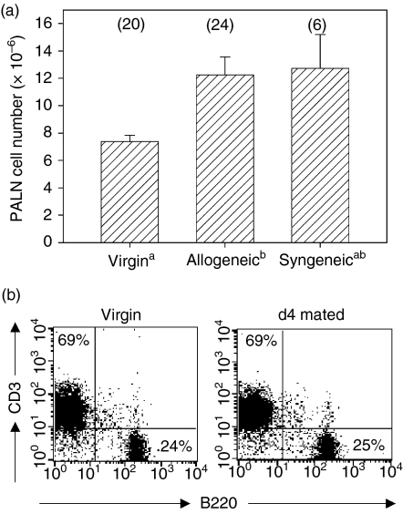
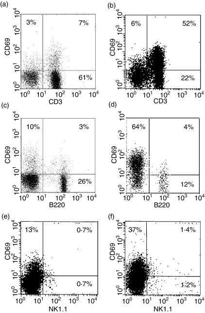
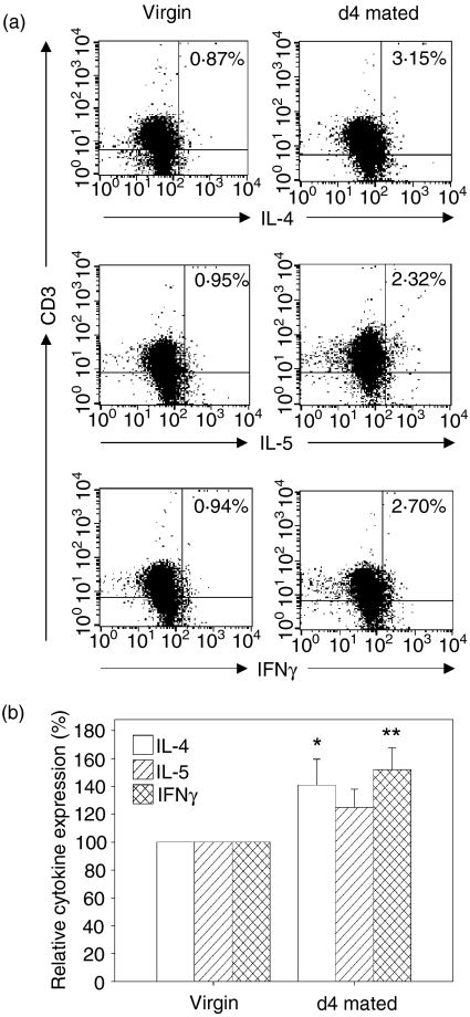
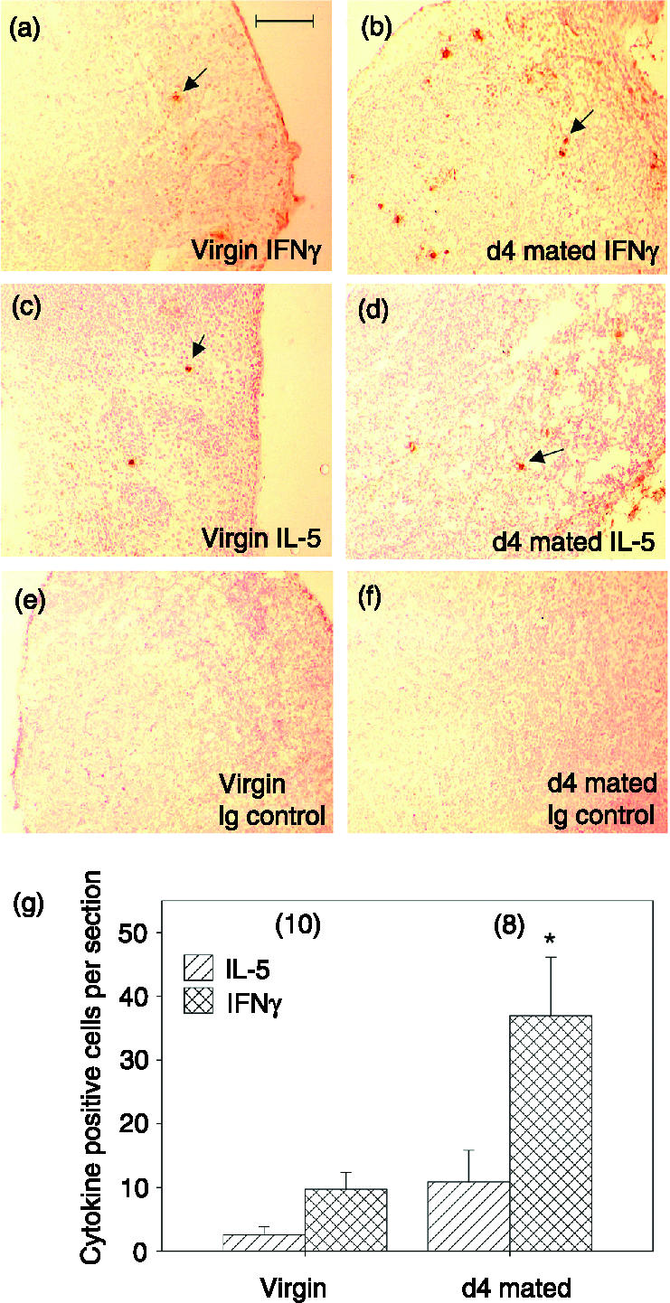
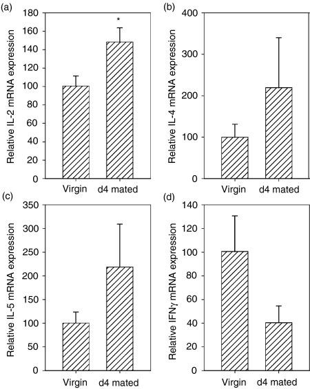
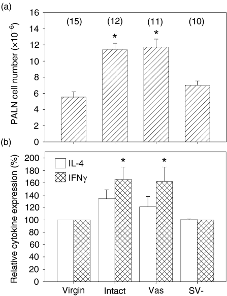
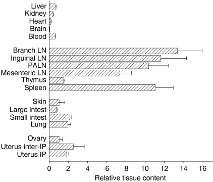
Insemination elicits inflammatory changes in female reproductive tissues, but whether this results in immunological priming to paternal antigens or influences pregnancy outcome is not clear. We have evaluated indices of lymphocyte activation in lymph nodes draining the uterus following allogeneic mating in mice and have investigated the significance of sperm and plasma constituents of semen in the response. At 4 days after mating, there was a 1b7-fold increase in the cellularity of the para-aortic lymph node (PALN) compared with virgin controls. PALN lymphocytes were principally T and B lymphocytes, with smaller populations of CD3(+) B220(lo), NK1.1(+) CD3(-) (NK) and NK1.1(+) CD3(+) (NKT) cells. CD69 expression indicative of activation was increased after mating and was most evident in CD3(+) and NK1.1(+) cells. Synthesis of cytokines including interleukin-2, interleukin-4 and interferon-gamma was elevated in CD3(+) PALN cells after exposure to semen, as assessed by intracellular cytokine fluorescence-activated cell sorting, immunohistochemistry and quantitative reverse transcriptase polymerase chain reaction. Matings with vasectomized males indicated that the lymphocyte activation occurs independently of sperm. However, in contrast, males from which seminal vesicle glands were surgically removed failed to stimulate PALN cell proliferation or cytokine synthesis. Adoptive transfer experiments using radiolabelled lymphocytes from mated mice showed that lymphocytes activated at insemination home to embryo implantation sites in the uterus as well as other mucosal tissues and lymph nodes. These findings indicate that activation and expansion of female lymphocyte populations occurs after mating, and is triggered by constituents of seminal plasma derived from the seminal vesicle glands. Moreover, lymphocytes activated at insemination may help mediate maternal tolerance of the conceptus in the implantation site.
Figures
Similar articles
-
Paternal antigen-specific proliferating regulatory T cells are increased in uterine-draining lymph nodes just before implantation and in pregnant uterus just after implantation by seminal plasma-priming in allogeneic mouse pregnancy.J Reprod Immunol. 2015 Apr;108:72-82. doi: 10.1016/j.jri.2015.02.005. Epub 2015 Mar 9. J Reprod Immunol. 2015. PMID: 25817463
-
Cross-presentation of male seminal fluid antigens elicits T cell activation to initiate the female immune response to pregnancy.J Immunol. 2009 Jun 15;182(12):8080-93. doi: 10.4049/jimmunol.0804018. J Immunol. 2009. PMID: 19494334
-
Seminal fluid regulates accumulation of FOXP3+ regulatory T cells in the preimplantation mouse uterus through expanding the FOXP3+ cell pool and CCL19-mediated recruitment.Biol Reprod. 2011 Aug;85(2):397-408. doi: 10.1095/biolreprod.110.088591. Epub 2011 Mar 9. Biol Reprod. 2011. PMID: 21389340
-
Post-mating inflammatory responses of the uterus.Reprod Domest Anim. 2012 Aug;47 Suppl 5:31-41. doi: 10.1111/j.1439-0531.2012.02120.x. Reprod Domest Anim. 2012. PMID: 22913558 Review.
-
Influence of semen on inflammatory modulators of embryo implantation.Soc Reprod Fertil Suppl. 2006;62:231-45. Soc Reprod Fertil Suppl. 2006. PMID: 16866321 Review.
Cited by
-
Extracellular vesicles in host-pathogen interactions and immune regulation - exosomes as emerging actors in the immunological theater of pregnancy.Heliyon. 2019 Aug 31;5(8):e02355. doi: 10.1016/j.heliyon.2019.e02355. eCollection 2019 Aug. Heliyon. 2019. PMID: 31592031 Free PMC article. Review.
-
The Role of Type I and Type II NKT Cells in Materno-Fetal Immunity.Biomedicines. 2021 Dec 14;9(12):1901. doi: 10.3390/biomedicines9121901. Biomedicines. 2021. PMID: 34944717 Free PMC article. Review.
-
Effect of Exposure to Seminal Plasma Through Natural Mating in Cattle on Conceptus Length and Gene Expression.Front Cell Dev Biol. 2020 May 12;8:341. doi: 10.3389/fcell.2020.00341. eCollection 2020. Front Cell Dev Biol. 2020. PMID: 32478076 Free PMC article.
-
Seminal vesicle fluid ameliorates autoimmune response within central nervous system.Cell Mol Immunol. 2015 Jan;12(1):116-8. doi: 10.1038/cmi.2014.88. Epub 2014 Sep 22. Cell Mol Immunol. 2015. PMID: 25242273 Free PMC article. No abstract available.
-
Alternative pathway activation in pregnancy, a measured amount "complements" a successful pregnancy, too much results in adverse events.Immunol Rev. 2023 Jan;313(1):298-319. doi: 10.1111/imr.13169. Epub 2022 Nov 15. Immunol Rev. 2023. PMID: 36377667 Free PMC article. Review.
References
-
- Hutter H, Dohr G. HLA expression on immature and mature human germ cells. J Reprod Immunol. 1998;38:101–22. - PubMed
-
- Alexander NJ, Anderson DJ. Immunology of semen. Fertil Steril. 1987;47:192–204. - PubMed
-
- Kelly RW, Critchley HO. Immunomodulation by human seminal plasma: a benefit for spermatozoon and pathogen? Hum Reprod. 1997;12:2200–7. - PubMed
-
- Beer AE, Billingham RE. Host responses to intra-uterine tissue, cellular and fetal allografts. J Reprod Fert Suppl. 1974;21:59–88.
-
- Robertson SA, Mau VJ, Tremellen KP, Seamark RF. Role of high molecular weight seminal vesicle proteins in eliciting the uterine inflammatory response to semen in mice. J Reprod Fertil. 1996;107:265–77. - PubMed
Publication types
MeSH terms
Substances
LinkOut - more resources
Full Text Sources
Research Materials