Clinical Trial
. 2004 Jun;112(2):328-38.
doi: 10.1111/j.1365-2567.2004.01877.x.

Modulation of immune responses to bovine herpesvirus-1 in cattle by immunization with a DNA vaccine encoding glycoprotein D as a fusion protein with bovine CD154

Affiliations
Clinical Trial

Modulation of immune responses to bovine herpesvirus-1 in cattle by immunization with a DNA vaccine encoding glycoprotein D as a fusion protein with bovine CD154

Sharmila Manoj et al. Immunology. 2004 Jun.

Abstract

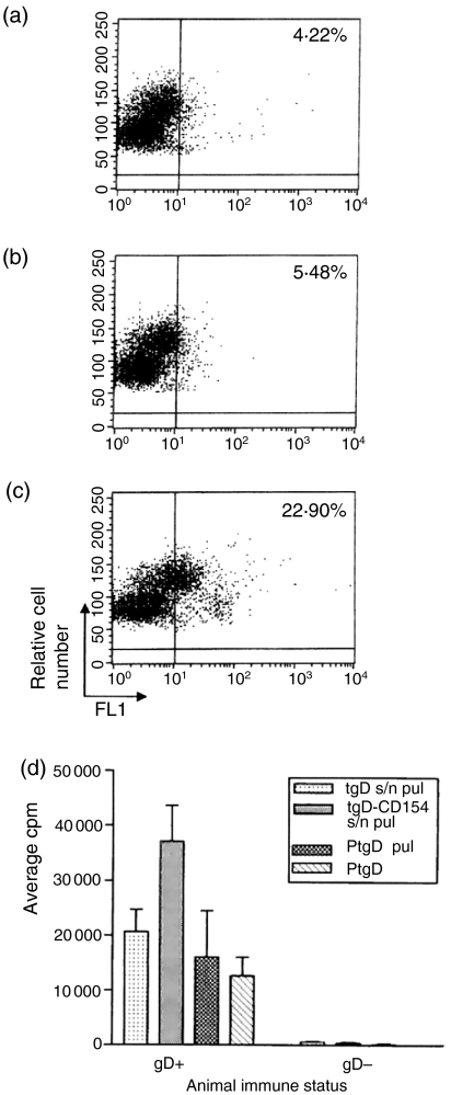
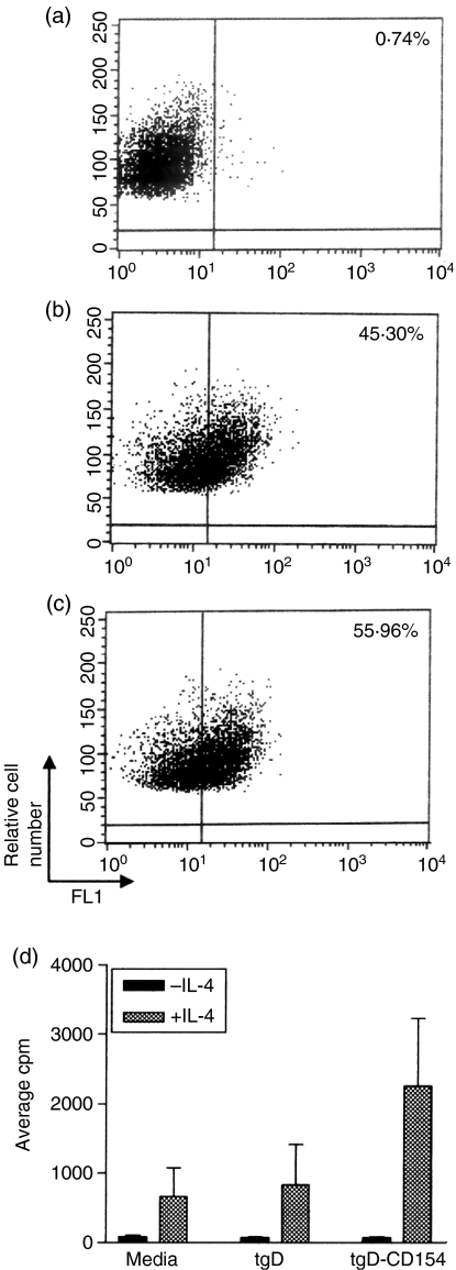
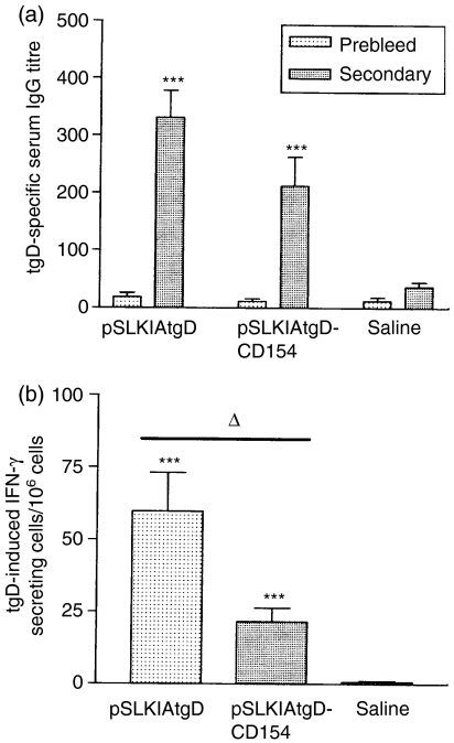
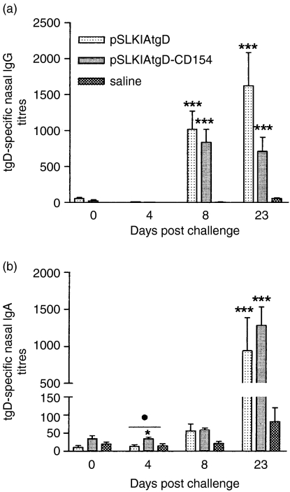
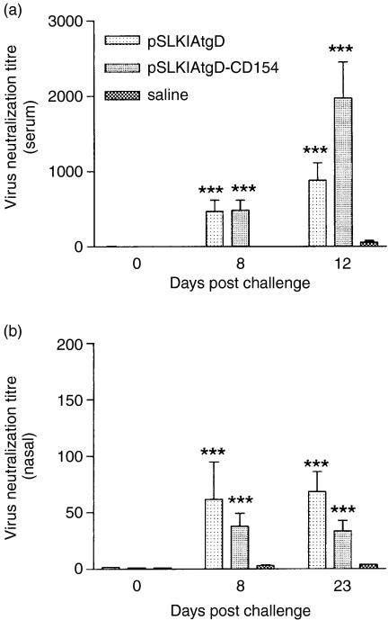
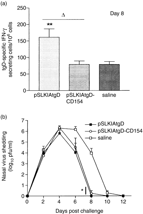
The objective of this study was to determine whether a DNA vaccine encoding bovine CD154 linked to a truncated version of bovine herpesvirus-1 (BHV-1) glycoprotein D (tgD-CD154) induces enhanced tgD-specific immune responses in cattle. In vitro characterization demonstrated that tgD and tgD-CD154 both bind to cultured bovine B cells, whereas only tgD-CD154 induces interleukin-4-dependent proliferation, suggesting that tgD-CD154 specifically binds the CD40 receptor and induces receptor signalling. Calves were immunized with plasmid encoding either tgD or tgD-CD154 by intradermal injection with a needle-free device. After two immunizations, tgD-specific immune responses were observed in both vaccinated groups and after challenge with BHV-1 these responses further increased. Animals immunized with plasmid encoding tgD-CD154 had significantly higher tgD-specific serum titres of immunoglobulins G and A but significantly lower numbers of tgD-specific interferon-gamma-secreting cells than animals immunized with plasmid encoding tgD after BHV-1 challenge. This suggests that the expression of an antigen as a chimeric protein with CD154 can qualitatively alter immune responses in cattle. Since we previously showed that plasmid encoding tgD-CD154 induces significantly enhanced secondary tgD-specific antibody responses in sheep, there appear to be interspecies differences in the immune responses induced by tgD-CD154, which suggests that both proteins in the chimeric molecule may influence protein targeting and the induction of an immune response.


Figures

Figure 1
Figure 1
Antigen-specific lymphocyte proliferation induced by binding of tgD or tgD-CD154 to bovine PBMCs. Supernatants from (a) mock-transfected, (b) pSLIAtgD-transfected, or (c) pSLIAtgD-CD154-transfected Cos-7 cells were incubated with bovine PBMCs. Bound protein was detected with a gD-specific mAb cocktail, followed by FITC-conjugated goat anti-mouse immunoglobulin. (d) PBMCs were isolated from naïve (tgD) and tgD immunized (tgD+) cattle (n=3) and incubated for 30 min at 4° with either pSLIAtgD (tgD s/n pul) or pSLIAtgD-CD154 (tgD-CD154 s/n pul) transfected Cos-7 cell supernatants or with 3 μg/well of purified tgD (PtgD pul). Subsequently, PBMCs were washed twice and 2 × 10⁵ cells/well were cultured for 72 hr. As a positive control, 2 × 10⁵ PBMCs/well were also cultured for 72 hr with 3 μg of purified tgD (PtgD). Cell proliferation was detected by adding methyl-[3H]thymidine during the last 8 hr of culture. Data are shown as average c.p.m.+SEM for triplicate cultures.
Figure 2
Figure 2
Induction of IL-4 responsiveness following tgD-CD154 binding to bovine B cells. Supernatants from (a) mock-transfected, (b) pSLIAtgD-transfected, or (c) pSLIAtgD-CD154-transfected Cos-7 cells were incubated with bovine B cells. Bound protein was detected with a gD-specific mAb cocktail, followed by FITC-conjugated goat anti-mouse immunoglobulin. (d) Triplicate cultures of bovine B cells (4 × 10⁴ cells/well) were incubated with medium or with pSLIAtgD- or pSLIAtgD-CD154 transfected Cos-7 cell supernatants. Cultures were incubated for 72 hr with either 100 μl/well of 10 ng/ml recombinant human IL-4 (+IL-4) or medium (–IL-4). Cell proliferation was detected by adding methyl-[3H]thymidine during the last 18 hr of culture. Data are shown as average c.p.m. + SEM for triplicate cultures.
Figure 3
Figure 3
Immune responses induced in calves by immunization with plasmids encoding tgD or tgD-CD154. Three groups (n=7) of calves were injected intradermally with saline, pSLKIAtgD or pSLKIAtgD-CD154. Responses were measured prior to primary immunization (prebleed) and 3 weeks post-secondary immunization (secondary). (a) tgD-specific mean IgG titres+SEM in sera. The OD reading (+2SD) corresponding to a known reciprocal dilution of standard positive control serum was used as the cut-off value to determine the ELISA titres. (b) Mean number of tgD-induced IFN-γ-secreting cells+SEM in blood. Values were calculated based on the difference between the number of spots per 106 cells in gD-stimulated wells and the number of spots per 106 cells in non-stimulated wells. Asterisks (***P<0·001) indicate significance of differences from the saline injected group. Open triangle (▵, P<0·05) indicates significance of difference between pSLKIAtgD- and pSLKIAtgD-CD154-immunized groups.
Figure 4
Figure 4
Antigen-specific IgG and IgA titres in sera of calves after BHV-1 challenge. Three groups (n = 7) of calves were injected intradermally with saline, pSLKIAtgD or pSLKIAtgD-CD154. Truncated gD-specific (a) mean IgG titres + SEM, (b) mean IgG1 and IgG2 titres+SEM and (c) mean IgA titres + SEM. The OD reading (+2SD) corresponding to a known reciprocal dilution of standard positive control serum was used as the cut-off value to determine the ELISA titres. Asterisks (***P < 0·001) indicate significance of differences from the saline injected group. Closed circle (•, P < 0·05) indicates significance of differences between the pSLKIAtgD- and pSLKIAtgD-immunized groups. Two animals injected with saline died on days 11 and 12 post-challenge.
Figure 5
Figure 5
Antigen-specific IgG and IgA titres in nasal secretions of calves following BHV-1 challenge. Three groups (n + 7) of calves were injected intradermally with saline, pSLKIAtgD or pSLKIAtgD-CD154. Truncated gD-specific (a) mean IgG titres + SEM, (b) mean IgA titres + SEM. The OD reading (+ 2SD) corresponding to a known reciprocal dilution of standard positive control serum was used as the cut-off value to determine the ELISA titres. Asterisks (***P < 0·01; *P < 0·05) indicate significance of differences from the saline injected group. Closed circle (•, P < 0·05) indicates significance of difference from the pSLKIAtgD-immunized group. Two animals injected with saline died on days 11 and 12 post-challenge.
Figure 6
Figure 6
Virus neutralization titres in sera and nasal secretions of cattle after BHV-1 challenge. Virus neutralization titres of (a) sera and (b) nasal secretions. Mean titres + SEM are expressed as the reciprocal of the highest dilution of serum that caused a 50% reduction in viral plaques relative to the virus control. Asterisks (***P<0·001) indicate significance of differences from the saline group. Two animals injected with saline died on days 11 and 12 post-challenge.
Figure 7
Figure 7
Cellular immune responses and virus shedding in cattle after BHV-1 challenge. Three groups (n = 7) of calves were injected intradermally with saline, pSLKIAtgD or pSLKIAtgD-CD154. (a) tgD-specific mean number of IFN-γ-secreting cells + SEM, 8 days post-challenge. Values were calculated based on the difference between the number of spots per 106 cells in gD-stimulated wells and the number of spots per 106 cells in non-stimulated wells. (b) Mean virus shedding + SEM in nasal secretions of calves challenged with BHV-1. Asterisks (**P < 0·01, *P < 0·05) indicate significance of differences with saline-injected group. Open triangle (▵, P < 0·05) indicates significance of differences between pSLKIAtgD- and pSLKIAtgD-CD154-immunized groups. Two animals injected with saline died on days 11 and 12 post-challenge.
Figure 8
Figure 8
A model to illustrate priming/induction of immune responses by tgD-CD154 upon DNA immunization.

References

    1. Saffery R, Choo KH. Strategies for engineering human chromosomes with therapeutic potential. J Gene Med. 2002;4:5–13. - PubMed
    1. Pachuk CJ, McCallus DE, Weiner DB, Satishchandran C. DNA vaccines – challenges in delivery. Curr Opin Mol Ther. 2000;2:188–98. - PubMed
    1. Babiuk LA, Pontarollo R, Babiuk S, Loehr B, van Drunen Littel-van den Hurk S. Induction of immune responses by DNA vaccines in large animals. Vaccine. 2003;21:649–58. - PubMed
    1. Pasquini S, Xiang Z, Wang Y, He Z, Deng H, Blaszczyk-Thurin M, Ertl HC. Cytokines and costimulatory molecules as genetic adjuvants. Immunol Cell Biol. 1997;75:397–401. - PubMed
    1. Boyle JS, Brady JL, Lew AM. Enhanced responses to a DNA vaccine encoding a fusion antigen that is directed to sites of immune induction. Nature. 1998;392:408–11. - PubMed

Publication types

MeSH terms

LinkOut - more resources