

A novel arginine substitution mutation in 1A domain and a novel 27 bp insertion mutation in 2B domain of keratin 12 gene associated with Meesmann's corneal dystrophy
- PMID: 15148206
- PMCID: PMC1772161
- DOI: 10.1136/bjo.2003.032870
A novel arginine substitution mutation in 1A domain and a novel 27 bp insertion mutation in 2B domain of keratin 12 gene associated with Meesmann's corneal dystrophy
Abstract
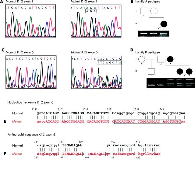
Aim: To determine the disease causing gene defects in two patients with Meesmann's corneal dystrophy.
Methods: Mutational analysis of domains 1A and 2B of the keratin 3 (K3) and keratin 12 (K12) genes from two patients with Meesmann's corneal dystrophy was performed by polymerase chain reaction amplification and direct sequencing.
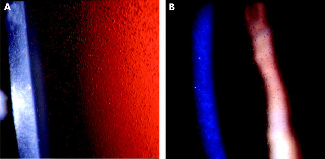
Results: Novel mutations of the K12 gene were identified in both patients. In one patient a heterozygous point mutation (429A-->C = Arg135Ser) was found in the 1A domain of the K12 gene. This mutation was confirmed by restriction digestion. In the second patient a heterozygous 27 bp duplication was found inserted in the 2B domain at nucleotide position 1222 (1222ins27) of the K12 gene. This mutation was confirmed by gel electrophoresis. The mutations were not present in unaffected controls.
Conclusion: Novel K12 mutations were linked to Meesmann's corneal dystrophy in two different patients. A missense mutation replacing a highly conserved arginine residue in the beginning of the helix initiation motif was found in one patient, and an insertion mutation, consisting of a duplication of 27 nucleotides, was found before the helix termination motif in the other.
Figures
Similar articles
-
Mutations in cornea-specific keratin K3 or K12 genes cause Meesmann's corneal dystrophy.Nat Genet. 1997 Jun;16(2):184-7. doi: 10.1038/ng0697-184. Nat Genet. 1997. PMID: 9171831
-
A novel keratin 12 mutation in a German kindred with Meesmann's corneal dystrophy.Br J Ophthalmol. 2000 May;84(5):527-30. doi: 10.1136/bjo.84.5.527. Br J Ophthalmol. 2000. PMID: 10781519 Free PMC article.
-
A novel mutation in KRT12 associated with Meesmann's epithelial corneal dystrophy.Br J Ophthalmol. 2002 Jul;86(7):729-32. doi: 10.1136/bjo.86.7.729. Br J Ophthalmol. 2002. PMID: 12084738 Free PMC article.
-
[Mutations in the keratin gene as a cause of Meesman-Wilke corneal dystrophy and autosomal dominant skin cornification disorders].Klin Monbl Augenheilkd. 2000 Jul;217(1):43-51. doi: 10.1055/s-2000-10382. Klin Monbl Augenheilkd. 2000. PMID: 10949816 Review. German.
-
The molecular genetics of keratin disorders.Am J Clin Dermatol. 2003;4(5):347-64. doi: 10.2165/00128071-200304050-00005. Am J Clin Dermatol. 2003. PMID: 12688839 Review.
Cited by
-
A novel mutation in the cornea-specific keratin 12 gene in Meesmann corneal dystrophy.Jpn J Ophthalmol. 2008 May-Jun;52(3):224-226. doi: 10.1007/s10384-007-0518-2. Epub 2008 Jul 27. Jpn J Ophthalmol. 2008. PMID: 18661274
-
Identification of presumed pathogenic KRT3 and KRT12 gene mutations associated with Meesmann corneal dystrophy.Mol Vis. 2015 Dec 31;21:1378-86. eCollection 2015. Mol Vis. 2015. PMID: 26788030 Free PMC article.
-
Identification of a novel mutation in the cornea specific keratin 12 gene causing Meesmann's corneal dystrophy in a German family.Mol Vis. 2010 May 29;16:954-60. Mol Vis. 2010. PMID: 20577595 Free PMC article.
-
Identification of a Novel Missense KRT12 Mutation in a Vietnamese Family with Meesmann Corneal Dystrophy.Case Rep Ophthalmol. 2020 Mar 17;11(1):120-126. doi: 10.1159/000506435. eCollection 2020 Jan-Apr. Case Rep Ophthalmol. 2020. PMID: 32308613 Free PMC article.
-
A Novel Pathogenic Variant in the KRT3 Gene in a Family with Meesmann Corneal Dystrophy.J Clin Med. 2025 Jan 28;14(3):851. doi: 10.3390/jcm14030851. J Clin Med. 2025. PMID: 39941522 Free PMC article.
References
-
- Irvine AD, Corden LD, Swensson O, et al. Mutations in cornea-specific keratin K3 or K12 genes cause Meesmann’s corneal dystrophy. Nat Genet 1997;16:184–7. - PubMed
-
- Meesmann A, Wilke F. Klinische und anatomische Untersuchungen uber eine bisher Unbekannte, dominant verebte Epitheldystrophie der Hornhaut. Klin Monatsbl Augenheilkd 1939:361–91.
-
- Fine BS, Yanoff M, Pitts E, et al. Meesmann’s epithelial dystrophy of the cornea. Am J Ophthalmol 1977;83:633–42. - PubMed
-
- Irvine AD, McLean WH. Human keratin diseases: the increasing spectrum of disease and subtlety of the phenotype-genotype correlation. Br J Dermatol 1999;140:815–28. - PubMed
-
- Lane EB. Keratins. In: Royce PM, Steinmann B, eds. Connective tissue and its heritable disorders. Molecular, genetic, and medical aspects. New York: Wiley-Liss, 1993:237–47.
Publication types
MeSH terms
Substances
LinkOut - more resources
Full Text Sources
Molecular Biology Databases