

Thyroid associated ophthalmopathy: evidence for CD4(+) gammadelta T cells; de novo differentiation of RFD7(+) macrophages, but not of RFD1(+) dendritic cells; and loss of gammadelta and alphabeta T cell receptor expression
- PMID: 15148216
- PMCID: PMC1772193
- DOI: 10.1136/bjo.2003.035915
Thyroid associated ophthalmopathy: evidence for CD4(+) gammadelta T cells; de novo differentiation of RFD7(+) macrophages, but not of RFD1(+) dendritic cells; and loss of gammadelta and alphabeta T cell receptor expression
Abstract
Aim: To characterise periorbital immune cells (stages, kinetics) in active and inactive thyroid associated ophthalmopathy (A-TAO; I-TAO).
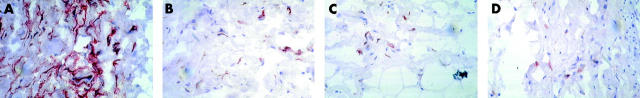
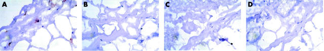
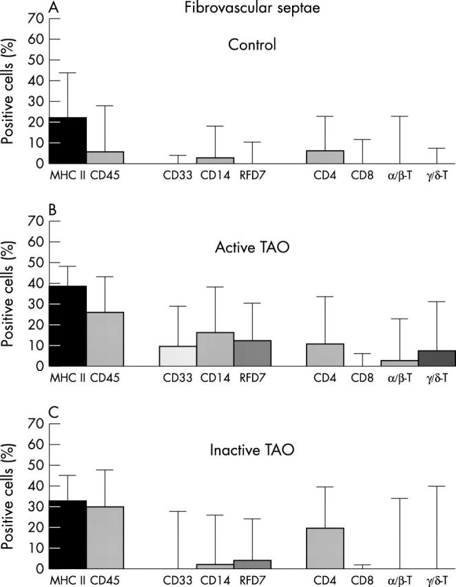
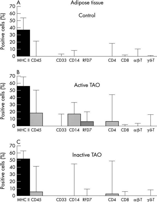
Methods: In orbital tissue cryosections of patients with A-TAO (n = 15), I-TAO (n = 11), and healthy controls (n = 14), adipose and fibrovascular areas were evaluated for MHC II(+) cells, CD45(+) total leukocytes, myeloid cells (CD33(+) monocytes; CD14(+) macrophages; mature RFD7(+) macrophages; RFD1(+) dendritic cells (DCs)), and lymphoid cells (CD4(+) T cells; alphabeta and gammadelta T cells; CD20(+) B cells). Results are expressed as medians and 5% confidence intervals.
Results: In fibrovascular septae, a surge of CD33(+) immigrants clearly correlating with disease activity generated significantly increased (p<0.05) percentages of CD14(+) and RFD7(+) macrophages. Intriguingly, CD4(+) cells were mostly gammadelta T cells, while alphabeta T helper cells were much less frequent. Successful treatment rendering TAO inactive apparently downregulates monocyte influx, macrophage differentiation, and T cell receptor expression. Similar trends were recorded for adipose tissue. Interestingly, RFD1(+) DCs were completely absent from all conditions examined.
Conclusion: A-TAO coincides with periorbital monocyte infiltration and de novo differentiation of macrophages, but not DCs. The authors discuss a novel potential role for inflammatory CD4(+) gammadelta T cells in TAO. Successful treatment apparently downregulates orbital monocyte recruitment and effects functional T cell knockout.
Figures
Similar articles
-
Discrimination of human macrophages and dendritic cells by means of monoclonal antibodies.Scand J Immunol. 1986 Sep;24(3):351-7. doi: 10.1111/j.1365-3083.1986.tb02104.x. Scand J Immunol. 1986. PMID: 3529367
-
Comparative gene expression by WC1+ gammadelta and CD4+ alphabeta T lymphocytes, which respond to Anaplasma marginale, demonstrates higher expression of chemokines and other myeloid cell-associated genes by WC1+ gammadelta T cells.J Leukoc Biol. 2006 Oct;80(4):939-52. doi: 10.1189/jlb.0506353. J Leukoc Biol. 2006. PMID: 17005908
-
T cells and fibroblasts in affected extraocular muscles in early and late thyroid associated ophthalmopathy.Br J Ophthalmol. 2000 May;84(5):517-22. doi: 10.1136/bjo.84.5.517. Br J Ophthalmol. 2000. PMID: 10781517 Free PMC article.
-
Monocytes and dendritic cells in a hypoxic environment: Spotlights on chemotaxis and migration.Immunobiology. 2008;213(9-10):733-49. doi: 10.1016/j.imbio.2008.07.031. Epub 2008 Sep 21. Immunobiology. 2008. PMID: 18926289 Review.
-
Orbital fibroblasts exhibit a novel pattern of responses to proinflammatory cytokines: potential basis for the pathogenesis of thyroid-associated ophthalmopathy.Thyroid. 2002 Mar;12(3):197-203. doi: 10.1089/105072502753600133. Thyroid. 2002. PMID: 11952039 Review.
Cited by
-
Graves' Ophthalmopathy on 68Ga-DOTANOC Positron Emission Tomography/Computed Tomography.Indian J Nucl Med. 2019 Oct-Dec;34(4):338-340. doi: 10.4103/ijnm.IJNM_147_19. Indian J Nucl Med. 2019. PMID: 31579195 Free PMC article.
-
Single-cell multiomic analysis unveils the immune landscape dynamics of graves' ophthalmopathy.Commun Biol. 2025 May 12;8(1):732. doi: 10.1038/s42003-025-08115-7. Commun Biol. 2025. PMID: 40355702 Free PMC article.
-
Cytokines in Thyroid-Associated Ophthalmopathy.J Immunol Res. 2022 Nov 14;2022:2528046. doi: 10.1155/2022/2528046. eCollection 2022. J Immunol Res. 2022. PMID: 36419958 Free PMC article. Review.
-
Molecular biomarkers of Graves' ophthalmopathy.Exp Mol Pathol. 2019 Feb;106:1-6. doi: 10.1016/j.yexmp.2018.11.004. Epub 2018 Nov 8. Exp Mol Pathol. 2019. PMID: 30414981 Free PMC article. Review.
-
Immunohistochemical analysis of human orbital tissue in Graves' orbitopathy.J Endocrinol Invest. 2020 Feb;43(2):123-137. doi: 10.1007/s40618-019-01116-4. Epub 2019 Sep 19. J Endocrinol Invest. 2020. PMID: 31538314 Review.
References
-
- Kazim M, Goldberg RA, Smith TJ. Insights into the pathogenesis of thyroid-associated orbitopathy: evolving rationale for therapy. Arch Ophthalmol 2002;120:380–6. - PubMed
-
- Wiersinga WM, Smit T, van der Gaag R, et al. Clinical presentation of Graves’ ophthalmopathy. Ophthalmic Res 1989;21:73–82. - PubMed
-
- Bahn RS, Heufelder AE. Pathogenesis of Graves’ ophthalmopathy. N Engl J Med 1993;329:1468–75. - PubMed
-
- Bartley GB, Fatourechi V, Kadrmas EF, et al. Clinical features of Graves’ ophthalmopathy in an incidence cohort. Am J Ophthalmol 1996;121:284–90. - PubMed
-
- Perros P, Kendall-Taylor P. Thyroid-associated ophthalmopathy: pathogenesis and clinical management. Baillieres Clin Endocrinol Metab 1995;9:115–35. - PubMed
Publication types
MeSH terms
Substances
LinkOut - more resources
Full Text Sources
Research Materials
Miscellaneous