

Sodium hyaluronate (hyaluronic acid) promotes migration of human corneal epithelial cells in vitro
- PMID: 15148219
- PMCID: PMC1772195
- DOI: 10.1136/bjo.2003.027573
Sodium hyaluronate (hyaluronic acid) promotes migration of human corneal epithelial cells in vitro
Abstract
Purpose: Sodium hyaluronate (hyaluronic acid) is known to promote corneal epithelial wound healing in vivo and in vitro, in animal experiments. Sodium hyaluronate is the ligand for CD44, a cell surface adhesion molecule which has been found on normal human corneal epithelial cells. The purpose of this study was to investigate the effect of sodium hyaluronate on human corneal epithelial cell migration, proliferation, and CD44 receptor expression.
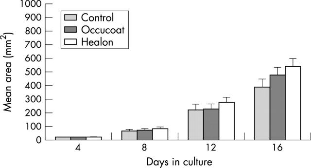
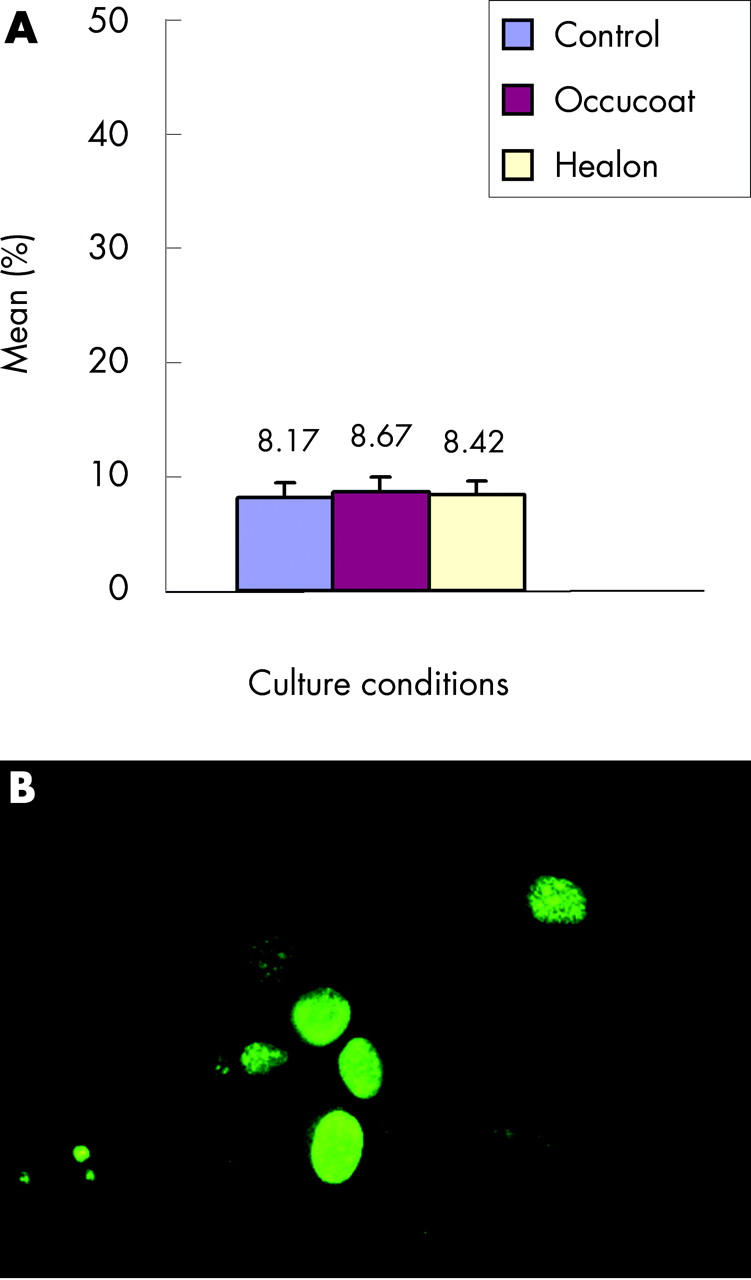
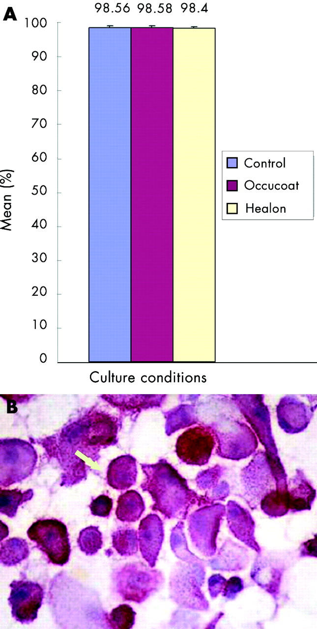
Methods: Human corneal epithelial cell cultures were established from 32 donor corneoscleral rims and maintained separately in three different culture conditions: (1) culture medium only, (2) sodium hyaluronate enriched (0.6 mg/ml) medium, and (3) hydroxypropylmethylcellulose enriched (2.5 mg/ml) medium. The total area of migrating epithelial cell sheets in each case was measured by planimetry on days 4, 8, 12, and 16. Cytospin preparations of cells cultured in the different culture conditions were examined immunohistochemically for proliferation and CD44 receptor expression using antibodies directed against Ki67 and CD44 respectively.
Results: Cells cultured in the presence of sodium hyaluronate showed significantly increased migration at days 12 and 16 (Friedmen test: p = 0.0012, day 16; p = <0.001, day 12) compared with cells cultured in the other media. There was no difference in cell proliferation (Ki67) or CD44 expression on cells cultured in the different culture conditions.
Conclusions: Sodium hyaluronate promotes migration but not proliferation or CD44 expression on human corneal epithelial cells in vitro. The beneficial effect of sodium hyaluronate in corneal wound healing is likely to be related to rapid migration of cells leading to rapid wound closure. This may be facilitated by the adhesion between CD44 on the cells and hyaluronic acid, which coats the surface of the denuded cornea.
Figures
References
-
- Dua HS, Forrester JV. The corneoscleral limbus in human corneal epithelial wound healing. Am J Ophthalmol 1990;110:646–56. - PubMed
-
- Dua HS, Forrester JV. Clinical patterns of corneal epithelial wound healing. Am J Ophthalmol 1987;104:481–9. - PubMed
-
- Inoue M, Katakami C. The effect of hyaluronic acid on corneal epithelial cell proliferation. Invest Ophthalmol Vis Sci 1993;34:13–15. - PubMed
-
- Koch DD, Liu JF, Glasser DB, et al. A comparison of corneal endothelial changes after use of healon or viscoat during phacoemulsification. Am J Ophthalmol 1993;115:188–201. - PubMed
MeSH terms
Substances
LinkOut - more resources
Full Text Sources
Other Literature Sources
Miscellaneous