Review
. 2004 Jun;88(6):844-7.
doi: 10.1136/bjo.2003.035584.

Treatment of Erdheim-Chester disease with cladribine: a rational approach

Review

Treatment of Erdheim-Chester disease with cladribine: a rational approach

C Myra et al. Br J Ophthalmol. 2004 Jun.
No abstract available


Figures

Figure 1
Figure 1
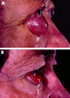
(A) The degree of bilateral proptosis and right chemosis in July 1999 in the patient with Erdheim-Chester disease, following treatment with cyclophosphamide. (B) The degree of proptosis and chemosis following treatment with cladribine.
Figure 2
Figure 2
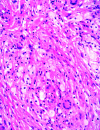
Biopsy of paravertebral tissue showing fibrocollagenous tissue, epithelioid cells, and Touton multinucleate giant cells. The biopsy was negative for S-100 protein.
Figure 3
Figure 3
X ray of long bones showing sclerosis and increased trabecular markings.
Figure 4
Figure 4
Serial monocyte counts in the patient with Erdheim-Chester disease (day 0 equivalent to 14 January 1999). The shaded area denotes the laboratory normal range (0.2–0.8×10⁹/l). Drug treatment periods are indicated by horizontal arrows (CPh = cyclophosphamide, Etop = etoposide, CyA = cyclosporine, and six courses of cladribine). Peripheral blood sampling for PCR based immunological analysis was perfomed on day 195.
Figure 5
Figure 5
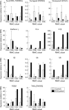
Quantitative RT-PCR analysis of cytokine expression on this patient with Erdheim-Chester disease (solid bars) compared to seven normal controls (open bars). A highly distinctive pattern of cytokine activation was found in the peripheral blood. Interleukin-1α (IL-1α), IL-1β, IL-2, and IL-8 all had raised expression compared with controls, consistent with monocyte activation. Smaller rises were seen in IL-6 and tumour necrosis factor α (TNF-α).

References

    1. Veyssier-Belot C, Cacoub P, Caparros-Lefebvre D, et al. Erdheim-Chester disease. Clinical and radiologic characteristics of 59 cases. Medicine 1996;75:157–69. - PubMed
    1. Alper MG, Zimmerman LE, Piana FG. Orbital manifestations of Erdheim-Chester disease. Trans Am Ophthalmol Soc 1983;81:64–85. - PMC - PubMed
    1. Wright RA, Hermann RC, Parisi JE. Neurological manifestations of Erdheim-Chester disease. J Neurol Neurosurg Psychiatry 1999;66:72–5. - PMC - PubMed
    1. Miller RL, Sheeler LR, Bauer TW, et al. Erdheim-Chester disease. Case report and review of the literature. Am J Med 1986;80:1230–6. - PubMed
    1. Rozenberg I, Wechsler J, Koenig F, et al. Erdheim-Chester disease presenting as malignant exophthalmos. Br J Radiol 1986;59:173–7. - PubMed