

Effects of chronic treatment with escitalopram or citalopram on extracellular 5-HT in the prefrontal cortex of rats: role of 5-HT1A receptors
- PMID: 15148253
- PMCID: PMC1574969
- DOI: 10.1038/sj.bjp.0705800
Effects of chronic treatment with escitalopram or citalopram on extracellular 5-HT in the prefrontal cortex of rats: role of 5-HT1A receptors
Abstract
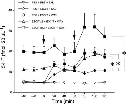
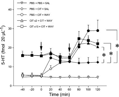
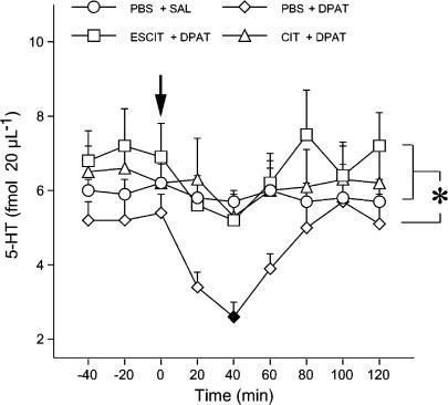
1 Microdialysis was used to study the acute and chronic effects of escitalopram (S-citalopram; ESCIT) and chronic citalopram (CIT), together with the 5-HT1A receptor antagonist WAY100,635 (N-[2-[methoxyphenyl)-1-piperazinyl]ethyl]-N-(2-pyridinyl) cyclohexane carboxamide trihydrochloride) and the 5-HT1A receptor agonist 8-hydroxy-2-(di-n-propylamino)tetralin (8-OH-DPAT), on extracellular 5-hydroxytryptamine (5-HT) levels in the rat prefrontal cortex. 2 Extracellular 5-HT rose to 234 and 298% of basal values after subcutaneous (s.c.) acute doses of 0.15 and 0.63 mg kg(-1) ESCIT. No further increase was observed at 2.5 mg kg(-1) ESCIT (290%). 3 The effect of 13-day s.c. infusion of 10 mg kg(-1) day(-1) ESCIT on extracellular 5-HT (422% of baseline) was greater than after 2 days (257% of baseline), whereas exposure to ESCIT was similar. In contrast, the increase in extracellular 5-HT induced by the infusion of CIT for 2 (306%) and 13 days (302%) was similar. However, brain and plasma levels of S-citalopram in rats infused with CIT for 13 days were lower than after 2 days. 4 Acute treatment with 2.5 mg kg(-1) ESCIT or 5 mg kg(-1) CIT raised extracellular 5-HT by 243 and 276%, respectively, in rats given chronic vehicle but had no effect in rats given ESCIT (10 mg kg(-1) day(-1)) or CIT (20 mg kg(-1) day(-1)) for 2 or 13 days, suggesting that the infused doses had maximally increased extracellular 5-HT. WAY100,635 (0.1 mg kg(-1) s.c.) increased extracellular 5-HT levels by 168, 174 and 169% of prechallenge values in rats infused with vehicle or ESCIT for 2 or 13 days, respectively. WAY100,635 enhanced extracellular 5-HT levels to 226, 153 and 164% of prechallenge values in rats infused with vehicle or CIT for 2 and 13 days, respectively. 5 8-OH-DPAT (0.025 mg kg(-1)) reduced extracellular 5-HT by 54% in control rats, but had no effect in those given ESCIT and CIT for 13 days. 6 This series of experiments led to the conclusion that chronic treatment with ESCIT desensitizes the 5-HT1A receptors, regulating the release of 5-HT in the prefrontal cortex and enhances the effect of the drug on extracellular 5-HT. They also indicate that chronic treatment with ESCIT and CIT did not prevent WAY100,635 from raising extracellular 5-HT.
Figures

References
-
- ADELL A., ARTIGAS F. Differential effects of clomipramine given locally or systemically on extracellular 5-hydroxytryptamine in raphe nuclei and frontal cortex. An in vivo brain microdialysis study. Naunyn Schmiedebergs Arch. Pharmacol. 1991;343:237–244. - PubMed
-
- ARBORELIUS L., NOMIKOS G.G., HERTEL P., SALMI P., GRILLNER P., HOOK B.B., HACKSELL U., SVENSSON T.H. The 5-HT1A receptor antagonist (S)-UH-301 augments the increase in extracellular concentrations of 5-HT in the frontal cortex produced by both acute and chronic treatment with citalopram. Naunyn Schmiedebergs Arch. Pharmacol. 1996;353:630–640. - PubMed
-
- ARTIGAS F., PEREZ V., ALVAREZ E. Pindolol induces a rapid improvement of depressed patients treated with serotonin reuptake inhibitors. Arch. Gen. Psychiatry. 1994;51:248–251. - PubMed
-
- AUERBACH S.B., HJORTH S. Effect of chronic administration of the selective serotonin (5-HT) uptake inhibitor citalopram on extracellular 5-HT and apparent autoreceptor sensitivity in rat forebrain in vivo. Naunyn Schmiedebergs Arch. Pharmacol. 1995;352:597–606. - PubMed
-
- BEL N., ARTIGAS F. Chronic treatment with fluvoxamine increases extracellular serotonin in frontal cortex but not in raphe nuclei. Synapse. 1993;15:243–245. - PubMed
Publication types
MeSH terms
Substances
LinkOut - more resources
Full Text Sources
