. 2004 May 24;165(4):493-503.
doi: 10.1083/jcb.200312171. Epub 2004 May 17.

c-Abl phosphorylates Dok1 to promote filopodia during cell spreading

Affiliations

c-Abl phosphorylates Dok1 to promote filopodia during cell spreading

Pamela J Woodring et al. J Cell Biol. .

Abstract

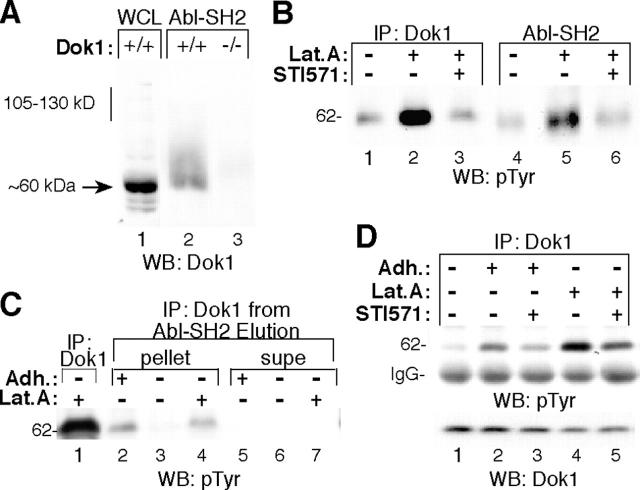
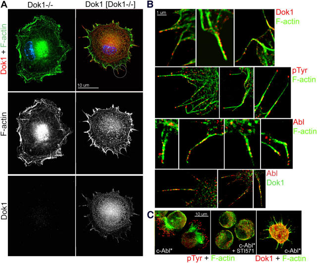
Filopodia are dynamic F-actin structures that cells use to explore their environment. c-Abl tyrosine kinase promotes filopodia during cell spreading through an unknown mechanism that does not require Cdc42 activity. Using an unbiased approach, we identified Dok1 as a specific c-Abl substrate in spreading fibroblasts. When activated by cell adhesion, c-Abl phosphorylates Y361 of Dok1, promoting its association with the Src homology 2 domain (SH2)/SH3 adaptor protein Nck. Each signaling component was critical for filopodia formation during cell spreading, as evidenced by the finding that mouse fibroblasts lacking c-Abl, Dok1, or Nck had fewer filopodia than cells reexpressing the product of the disrupted gene. Dok1 and c-Abl stimulated filopodia in a mutually interdependent manner, indicating that they function in the same signaling pathway. Dok1 and c-Abl were both detected in filopodia of spreading cells, and therefore may act locally to modulate actin. Our data suggest a novel pathway by which c-Abl transduces signals to the actin cytoskeleton through phosphorylating Dok1 Y361 and recruiting Nck.


Figures

Figure 1.
Figure 1.
pTyr-containing proteins associate with Abl-SH2 during FN-stimulated cell spreading. (A) NIH3T3 fibroblasts were detached from the substratum and held in suspension for 45–60 min, and then half of the cells were plated onto FN for 20 min (Adh.: +) while the other half remained in suspension (Adh.: −). Cell lysates were incubated with GST-agarose (lane 1) or GST-Abl-SH2-agarose (lanes 2 and 3). After washing the beads stringently, bound proteins were subjected to Western blot analysis using pTyr antibodies (WB:pTyr). (B) Abl−/−Arg−/− MEFs stably reconstituted with vector (pMSCV, lanes 1 and 2) or c-Abl (lanes 3 and 4) were pretreated with 5 μM STI571 (lanes 2 and 4) or vehicle (DMSO, lanes 1 and 3) for 8 h. Cell lysates were incubated with Abl-SH2 and processed as in A. (C and D) Abl[Abl−/−Arg−/−] MEFs were maintained in suspension for 60 min (C) or replated onto FN (D). As indicated, 1 μM latrunculin A (Lat. A), 1 μM STI571, or 0.5 μM SU6656 was added to culture media before cell detachment (C, lanes 2–4). Cell lysates were incubated with Abl-SH2 and processed as described in A.
Figure 2.
Figure 2.
Dok1 is tyrosine phosphorylated when Abl is active in cells. MEFs were pretreated with STI571 and latrunculin A as indicated (see Materials and methods), and then detached and either held in suspension (Adh.: −) or replated onto FN for 20 min (Adh.: +). (A) Lysates from latrunculin-treated passage 3 MEFs null for Dok1 (Dok1−/−) or littermate control MEFs (Dok1+/+) were incubated with Abl-SH2, and the bound Dok1 was detected using Dok1 antibodies in Western blot analysis. Lane 1 contained whole cell lysate as a positional control. (B) Dok1 was immunoprecipitated (α6043) from the same suspended cell lysates (lanes 1–3) as used for the Abl-SH2 affinity analysis (lanes 4–6). Precipitated proteins were subjected to Western blot analysis using pTyr antibodies. Note that the slight variation in mobility of p62 between the immunoprecipitates and the Abl-SH2 pull downs is likely due to the presence of a large quantity of Abl-SH2 domain in lanes 4–6. (C) Abl-SH2 was incubated with the indicated cell lysates, and the bound pTyr-containing proteins were eluted from the agarose with 0.5 M phenylphosphate. Eluates were diluted and subjected to immunoprecipitation with Dok1 antibodies. Equal fractions from the supernatant (lanes 5–7) and pellet (lanes 2–4) of the Dok1 immunoprecipitate were subjected to Western blot analysis using pTyr antibodies. Lane 1 contained a Dok1 immunoprecipitate from latrunculin-treated cells. (D) Dok1 was immunoprecipitated from lysates of the indicated Abl[Abl−/−Arg−/−] MEFs, and then subjected to sequential Western blot analysis using pTyr antibodies, followed by Dok1 antibodies.
Figure 3.
Figure 3.
Expression of c-Abl increases the pTyr on Dok1 but not Y361F Dok1. (A) HA-Dok1 mutants were expressed stably in Abl−/−Arg−/− MEFs previously reconstituted with either pMSCV empty vector (lane 1) or c-Abl (lanes 2–7). The mutated tyrosines (Y to F) in HA-Dok1 are indicated at the top of each lane. HA-Dok1 mutants were immunoprecipitated from cell lysates of spreading cells and subjected to sequential Western blot analysis using pTyr antibodies, and then Dok1 antibodies. (B) Tryptic phosphopeptide maps of GST-Dok1 and GST-Y361F Dok1. Purified GST-Dok1 proteins were phosphorylated using γ-[32P]ATP and purified c-Abl. GST-Dok1 was isolated with glutathione-Sepharose and subjected to SDS-PAGE. The 32P-labeled Dok1 band was excised from the gel and processed for tryptic phosphopeptide mapping (see Materials and methods). The sample origin is located at the lower left. Circles drawn on the right panel indicate where peptides 1 and 2 would have run if they were present in the map of Y361F Dok1. (C) Peptide 1 was purified from the TLC plate of WT 32P-Dok1 shown in B. The map of purified peptide 1 incubated in buffer without trypsin (top) and the map of purified peptide 1 redigested with trypsin (bottom) are shown. (D) The indicated Abl[Abl−/−Arg−/−] MEFs described in A were held in suspension for 45–60 min, and then half of the cells were plated onto FN for 20 min (lanes 2 and 4), while the other half remained in suspension (lanes 1 and 3). HA-Dok1 constructs were immunoprecipitated from cell lysates and subjected to sequential Western blot analysis using pTyr antibodies, and then Dok1 antibodies.
Figure 4.
Figure 4.
c-Abl increases the phosphorylation of Y361 of Dok1 in cells. (A) Tryptic phosphopeptide map of HA-Dok1 from 32P-labeled HEK293T cells that were coexpressing c-Abl. HA-Dok1 was immunoprecipitated and processed for tryptic phosphopeptide mapping. Before spotting onto the TLC plate, trypsin-digested synthetic pY361 Dok1 peptides (see Materials and methods) were added to the 32P-Dok1 sample. Arrows indicate the position of synthetic pY361 Dok1 peptides detected by ninhydrin staining. The autoradiogram (left) was obtained by exposing the ninhydrin-stained TLC plate (right) to film. The mobility of the synthetic pY361 Dok1 peptides corresponds to that of the phosphopeptides labeled “1” and “2” derived from 32P-Dok1 in Fig. 3 B. (B) Phosphoamino acid analysis of WT Dok1 and Y361F Dok1 from 32P-labeled HEK293T cells that were coexpressing c-Abl. Samples were prepared as described previously (Meisenhelder et al., 1999). Right panel shows ninhydrin staining indicating the position of the phosphoamino acid standards that were added to the 32P samples. (C) Dok1 was immunoprecipitated from lysates of the indicated cells, and then subjected to Western blot analysis using the antibodies indicated at the far right of each row of panels. (left) HA-Dok1 was immunoprecipitated from lysates of HEK293T cells that were transfected with empty vector (lane 1); KD Abl and WT Dok1 (lane 2); WT c-Abl and WT Dok1 (lane 3); or WT c-Abl and Y361F Dok1 (lane 4). (bottom left) pY361-Dok1 synthetic peptide 1 was added to the to the αpY361Dok1 immunoblotting solution. (middle) HA-Dok1 was immunoprecipitated from lysates of Abl[Abl−/−Arg−/−] MEFs expressing either WT Dok1 (lanes 1–3) or Y361F Dok1 (lane 4). MEFs were held in suspension (lane 1), replated on FN (lanes 2 and 4), or pretreated with STI571 and replated on FN (lane 3). (right) Endogenous Dok1 was immunoprecipitated (α6043) from the following spreading MEFs: lane 1, Dok1−/−; lane 2, Abl[Abl−/−Arg−/−]; or lane 3, pMSCV[Abl−/−Arg−/−]. Because endogenous Dok1 migrates close to the IgG heavy chain, Dok1−/− MEFs were used as a control (lane 1). Note that the intensity level of signals between the left, middle, and right panels are not directly comparable because these results are from three separate experiments.
Figure 5.
Figure 5.
Stable expression of activated c-Abl (c-Abl*) in nonadherent fibroblasts increases the pTyr content on Dok1 pulled down with Abl-SH2. (A) c-Abl or activated c-Abl* (c-Abl missing the last four amino acids; Woodring et al., 2002) was stably expressed in Abl−/− MEFs. The deconvolution microscopy images shown highlight the differences observed in cells that were fixed after 60 min in suspension (Woodring et al., 2002). The F-actin cytoskeleton was stained using TRITC-phalloidin. (B) Cell lysates from FN-replated (lanes 1 and 3) and suspended (lanes 2 and 4) MEFs were incubated with Abl-SH2, and the bound proteins were subjected to Western blot analysis using pTyr antibodies. Arrow indicates the p62 kD band we identified as Dok1 (Fig. 2).
Figure 6.
Figure 6.
Dok1 expression increases Abl-dependent filopodia during cell spreading, whereas Y361F Dok1 expression does not affect filopodia. (A) The indicated Abl−/−Arg−/− MEF lines were held in suspension for 45–60 min, and then replated onto FN-coated coverslips for 20 min before fixation. Fixed cells were stained with TRITC-phalloidin, and images were obtained using deconvolution microscopy. Images shown highlight the differences between the populations of cells. (B) The number of F-actin microspikes/filopodia were counted on 200–1,000 randomly selected cells from each indicated cell line as described in Materials and methods. Error bars represent SEM; *, P < 0.001 compared with the parent cell line not expressing Dok1.
Figure 7.
Figure 7.
Dok1 / primary MEFs have fewer filopodia. Spreading passage 3 Dok1−/− MEFs (bottom row, middle and right) and the littermate Dok1+/+ MEFs (top row, middle and right) were analyzed side-by-side with spreading Abl[Abl−/−Arg−/−] and pMSCV[Abl−/− Arg−/−] MEFs (left panels, top and bottom, respectively). TRITC-phalloidin staining (A), and quantification of the number of F-actin microspikes/filopodia in spreading cells and statistics (B and C) were performed as described in Fig. 6. Error bars represent SEM; *, P < 0.001 when the values (represented by the columns) on either side of the horizontal line are compared. (C) As indicated, MEFs were pretreated with STI571 or were stably infected with c-Abl or control (pMSCV) retrovirus and selected for 4 d. Note that data for the histograms was normalized to WT MEFs (set to 1) so that experiments performed on separate occasions could be compared.
Figure 8.
Figure 8.
Dok1, pTyr, and c-Abl localize to the tips of filopodia and peripheral actin microspikes. (A) Passage 5 Dok1−/− MEFs were infected with pBABE-puro vector (left) or Dok1 retrovirus (right), and then selected for 4 d. MEFs were fixed during spreading on FN (20 min) and stained. FITC-phalloidin was used to detect F-actin (green); Dok1 (A3) + Texas red goat–αmouse antibodies were used to detect Dok1 (red); and Hoechst 33258 (blue) was used to detect nuclei. (B) Spreading MEFs were stained with FITC-phalloidin and the following antibodies: Dok1 (A3, first row), pTyr (4G10, second row), or c-Abl (8E9, third row). Texas red goat–αmouse antibodies were used as secondary antibodies. Abl, Dok costaining (fourth row) was performed sequentially, first staining with αDok (A3) + goat αmouse-Alexa Fluor 488, then with αAbl (8E9, mouse mAb) directly conjugated to Alexa Fluor 546. All panels show images of filopodia protruding from MEFs spreading on FN. The top left panel is a higher magnification of the circled area in A. (C, left and middle) FITC-phalloidin and αpTyr staining of suspended cells stably expressing c-Abl*, which remains active in suspended cells. Cells were pretreated with STI571 as indicated. (right) FITC-phalloidin and αDok1 (A3) staining of a suspended cell stably expressing c-Abl*.
Figure 9.
Figure 9.
The Nck adaptor protein is involved in Abl-dependent filopodia formation. (A and B) Abl−/−Arg−/− MEFs were stably infected with pMSCV empty vector, c-Abl, WT Dok1, and/or Y361F Dok1 retrovirus as indicated. MEFs were held in suspension for 60 min, and then lysed (B, lane 9) or replated onto FN for 25 min (all other lanes in A and B). (A) Endogenous Nck and Dok1 were immunoprecipitated with α5547 (lanes 1–3) and α12CA5 (lanes 4 and 5), respectively. Western blot analysis was performed using antibodies indicated at the right. (B) Lanes 1–3 contain whole cell lysates, and lanes 4–10 contain Nck immunoprecipitates from lysates of Abl[Abl−/−Arg−/−] MEFs expressing endogenous Dok1 (lanes 3 and 6–8) or increased levels of Dok1 (lanes 1, 2, 4, 5, 9, and 10). As indicated, MEFs were pretreated with DMSO (lane 7) or 5 μM STI571 (lane 8). Exposure time for lanes 7 and 8 was ∼10 times longer than for lanes 4–6. (C and D) MEFs null for Nck1 and Nck2 were stably reconstituted with empty vector (left) or Nck1 (right). Cells were plated on FN for 25 min, fixed, and stained with TRITC-phalloidin. The number of F-actin microspikes and filopodia were counted as described in Materials and methods. *, P < 0.001 compared with the cell line lacking Nck.

References

    1. Benesch, S., S. Lommel, A. Steffen, T. Stradal, N. Scaplehorn, M. Way, J. Wehland, and K. Rottner. 2002. PIP2-induced vesicle movement depends on N-WASP and involves Nck, WIP and Grb2. J. Biol. Chem. 277:37771–37776. - PubMed
    1. Blake, R., M. Broome, X. Liu, J. Wu, M. Gishizky, L. Sun, and S. Courtneidge. 2000. SU6656, a selective src family kinase inhibitor, used to probe growth factor signaling. Mol. Cell. Biol. 20:9018–9027. - PMC - PubMed
    1. Campellone, K., S. Rankin, T. Pawson, M. Kirschner, D. Tipper, and J. Leong. 2004. Clustering of Nck by a 12-residue Tir phosphopeptide is sufficient to trigger localized actin assembly. J. Cell Biol. 164:407–416. - PMC - PubMed
    1. Carpino, N., D. Wisniewski, A. Strife, D. Marshak, R. Kobayashi, B. Stillman, and B. Clarkson. 1997. p62(Dok): a constitutively tyrosine-phosphorylated, GAP-associated protein in chronic myelogenous leukemia progenitor cells. Cell. 88:197–204. - PubMed
    1. Cong, F., B. Yuan, and S. Goff. 1999. Characterization of a novel member of the DOK family that binds and modulates v-Abl signaling. Mol. Cell. Biol. 19:8314–8325. - PMC - PubMed

Publication types

MeSH terms