. 2004 May 17;199(10):1355-65.
doi: 10.1084/jem.20032196.

T cell-specific ablation of Fas leads to Fas ligand-mediated lymphocyte depletion and inflammatory pulmonary fibrosis

Affiliations

T cell-specific ablation of Fas leads to Fas ligand-mediated lymphocyte depletion and inflammatory pulmonary fibrosis

Zhenyue Hao et al. J Exp Med. .

Abstract

To study the role of Fas-Fas ligand (FasL) interaction-mediated apoptosis in lymphocyte homeostasis, we generated a mutant fas allele allowing conditional inactivation of the fas gene through Cre-mediated recombination. Experiments in which Fas was ablated in T cells, B cells, T and B cells, or in a more generalized manner demonstrated that the development of lymphoproliferative disease as seen in Fas-deficient mice requires Fas ablation in lymphoid and nonlymphoid tissues. Selective inactivation of Fas in T cells led to a severe lymphopenia over time, accompanied by up-regulation of FasL on activated T cells and apoptosis of peripheral lymphocytes. In addition, the mutant animals developed a fatal wasting syndrome caused by massive leukocyte infiltration in the lungs together with increased inflammatory cytokine production and pulmonary fibrosis. Inhibition of Fas-FasL interaction in vivo completely prevented the loss of lymphocytes and initial lymphocyte infiltration in the lungs. Thus, FasL-mediated interaction of activated, Fas-deficient T cells with Fas-expressing cells in their environment leads to break down of lymphocyte homeostasis and development of a lung disease strikingly resembling idiopathic pulmonary fibrosis in humans, a common and severe disease for which the mutant mice may serve as a first animal model.


Figures
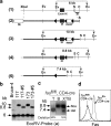
Figure 1.
Figure 1.
T cell–specific KO of Fas. (a) Targeting scheme. (1) Genomic structure of the fas locus surrounding exon IX. (2) Targeting vector construction. The chromosomal locus after homologous recombination is shown in 3. Cre-mediated deletion produces the fas floxed (4, fas fl) and deleted alleles (5, fas del). (triangles) loxP sites. Ev, EcoRV; S, SphI; C, ClaI restriction site. (b) Southern blot analysis on DNA from wild-type embryonic stem cells (Bruce-4) and homologous recombinants (clones 117 and 173 and neo r deleted clones 117#5 and 173#5). (c) T cell–specific deletion of Fas. T, thymocytes; B, purified splenic B cells (CD19+); XTB, purified nonlymphocyte population. Deletion efficiencies are indicated. (d) FACS® analysis showing loss of Fas expression in thymocytes of fas fl/fl, CD4-cre mice.
Figure 2.
Figure 2.
Defective Fas expression in nonlymphoid cells is required for the development of lymphoproliferative disease. FACS® analysis of DN T cells in LNs is shown. (a) 7–9-mo-old C57BL/6 mice with Fas deletion in tissue-specific or inducible manner. Mx–cre-mediated deletion was induced 8 mo before analysis. (b) Mild lymphadenopathy and splenomegaly upon T, B, and T plus B cell–specific Fas inactivation in (C57BL/6×MRL) F1 mice at the age of 5–6 mo. Pictures of spleen and LNs are shown. Weights are shown as means ± SD (n = 2∼6). Cell numbers shown on top of FACS® plots indicate total cellularity from the largest LNs of mice suffering from lymphadenopathy or of inguinal or cervical LNs of mice without apparent lymphadenopathy.
Figure 3.
Figure 3.
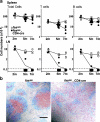
Loss of peripheral lymphocytes upon T cell–specific Fas inactivation. (a) Cellularity in the spleen and inguinal LNs. Each circle represents an individual mouse. Lines connect mean values of each group. (b) Immunohistochemical staining of spleens of 5–6-mo-old mice (four mice per group analyzed) with anti-CD3 (red) and anti-CD19 (blue) antibody. (c) Annexin V staining of T and B cells in LNs (four mice per group analyzed) at the age of 16 wk. (d) TUNEL staining of the splenic sections from animals (four mice per group analyzed) at the age of 16 wk. Green and red staining shows apoptotic cells and nuclei, respectively. (b and d) Bars, 200 μm.
Figure 3.
Figure 3.
Loss of peripheral lymphocytes upon T cell–specific Fas inactivation. (a) Cellularity in the spleen and inguinal LNs. Each circle represents an individual mouse. Lines connect mean values of each group. (b) Immunohistochemical staining of spleens of 5–6-mo-old mice (four mice per group analyzed) with anti-CD3 (red) and anti-CD19 (blue) antibody. (c) Annexin V staining of T and B cells in LNs (four mice per group analyzed) at the age of 16 wk. (d) TUNEL staining of the splenic sections from animals (four mice per group analyzed) at the age of 16 wk. Green and red staining shows apoptotic cells and nuclei, respectively. (b and d) Bars, 200 μm.
Figure 4.
Figure 4.
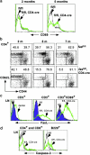
Activation and high levels of FasL expression in Fas-deficient T cells. (a) Expression of the activation marker CD69 on splenic CD3+ T cells. (b) Analysis of CD62L versus CD44 on gated CD4+ splenic cells to determine the proportion of naive (CD62Lhigh/CD44low) and activated/memory (CD62Llow/CD44high) T cells. (c) FasL expression on gated CD3+ or CD3+CD69+ LN cells from 5-mo-old animals. (d) Active caspase-3 activity in T and B cells of LN cells from 18-wk-old mice. Three to five mice per group were analyzed, and a representative analysis is shown for each group.
Figure 5.
Figure 5.
Wasting syndrome and increased inflammatory cytokine production in aged T cell–specific Fas-deficient mice. (a) A mutant and a control mouse at the age of 15 mo. (b) Survival of mutant and control animals. (c) MIP-2 and TGF-β1 levels in the BALF at the age of 14∼18 mo. Results shown are means ± SD (n = 3∼5).
Figure 6.
Figure 6.
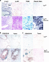
Leukocyte infiltration in the lungs and development of pulmonary fibrosis upon T cell–specific Fas inactivation. (a) Histological analysis of the lungs of mice at the age of 10 mo. H & E, hematoxylin and eosin staining; Ly-6G, staining for neutrophils (red); F4/80, staining for macrophages (blue); Elastic Stain, staining of elastic fibers (black blue) and cell nuclei (black). (b) Immunohistochemical staining of the lungs of 5–6-mo-old mice with anti-CD3 (red) and anti-CD19 (blue) antibody, and TUNEL (brown) on serial lung sections. (c) The staining was the same as in b, except the sections were from mice at the age of 15 mo. Three mice per group were analyzed, and representative results are shown. Bar, 100 μm.
Figure 7.
Figure 7.
Prevention of lymphopenia and cell infiltration in the lungs upon application of anti-FasL antibody. Groups of three mutant and three control mice were treated with anti-FasL antibody or isotype-matched hamster IgG1. Two mice from each group were analyzed 8 wk after antibody application, and one mouse from each group was analyzed at 12 wk. (a) FACS® analysis of lymphocytes in inguinal LNs. (b and c) Coimmunostaining of the spleen (b) and lung (c) sections with anti-CD3 (red) and anti-CD19 (blue) antibody. Shown is the analysis of one set of mice treated with antibody for 8 wk. (b and c) Bars, 200 μm.

References

    1. Krammer, P.H. 1999. CD95(APO-1/Fas)-mediated apoptosis: live and let die. Adv. Immunol. 71:163–210. - PubMed
    1. Krammer, P.H. 2000. CD95's deadly mission in the immune system. Nature. 407:789–795. - PubMed
    1. Watanabe-Fukunaga, R., C.I. Brannan, N.G. Copeland, N.A. Jenkins, and S. Nagata. 1992. Lymphoproliferation disorder in mice explained by defects in Fas antigen that mediates apoptosis. Nature. 356:314–317. - PubMed
    1. Takahashi, T., M. Tanaka, C.I. Brannan, N.A. Jenkins, N.G. Copeland, T. Suda, and S. Nagata. 1994. Generalized lymphoproliferative disease in mice, caused by a point mutation in the Fas ligand. Cell. 76:969–976. - PubMed
    1. Rieux-Laucat, F., F. Le Deist, C. Hivroz, I.A. Roberts, K.M. Debatin, A. Fischer, and J.P. de Villartay. 1995. Mutations in Fas associated with human lymphoproliferative syndrome and autoimmunity. Science. 268:1347–1349. - PubMed

Publication types