. 2004 May 17;199(10):1401-8.
doi: 10.1084/jem.20040249.

In vivo instruction of suppressor commitment in naive T cells

Affiliations

In vivo instruction of suppressor commitment in naive T cells

Irina Apostolou et al. J Exp Med. .

Abstract

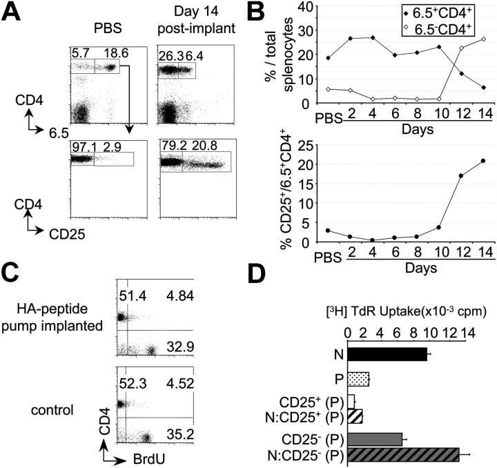
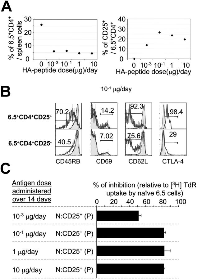
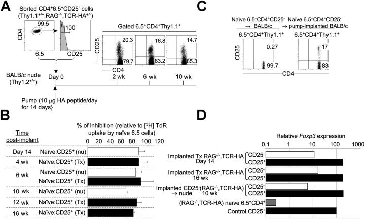
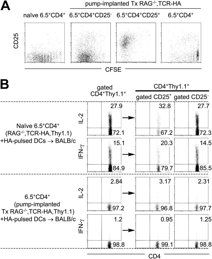
The induction of antigen-specific tolerance in the mature immune system of the intact organism has met with limited success. Therefore, nonspecific immunosuppression has been the treatment of choice to prevent unwanted immunity. Here, it is shown that prolonged subcutaneous infusion of low doses of peptide by means of osmotic pumps transforms mature T cells into CD4+25+ suppressor cells that can persist for long periods of time in the absence of antigen and confer specific immunologic tolerance upon challenge with antigen. The described procedure resembles approaches of tolerance induction used decades ago, induces tolerance in the absence of immunity, and holds the promise to become an effective means of inducing antigen-specific tolerance prospectively, whereas its power to suppress already ongoing immune responses remains to be determined.


Figures

Figure 1.
Figure 1.
Generation of CD25+ Ag-specific suppressor T cells by continuous peptide delivery for 14 d. (A) CD25 expression by 6.5+CD4+ splenocytes of Tx TCR-HA,RAG-2−/− mice 14 d after implantation of osmotic pumps delivering either PBS or 10 μg of HA peptide per day. (B) Percentages of 6.5+ or 6.5CD4+ (top) and of CD25+6.5+CD4+ (bottom) T cells at various days of peptide delivery or 14 d after PBS infusion. (C) BrdU incorporation by splenocytes of either Tx TCR-HA,RAG-2−/− mice within the 14 d of peptide osmotic pump infusion or control TCR-HA,RAG-2−/− mice. (D) In vitro proliferation of flow cytometry–purified CD4+6.5+ T cells from untreated TCR-HA,RAG-2−/−(N) or from Tx TCR-HA,RAG-2−/− mice infused with 10 μg/d of HA peptide for 14 d (P; black and dotted bars, respectively), as well as CD25+ (white bar) and CD25 (gray bar) subsets of 6.5+CD4+ cells from peptide-infused mice either cultured alone (unstriped bars) or cocultured with naive 6.5+CD4+ T cells from untreated TCR-HA,RAG-2−/− mice (striped bars) in the presence of HA peptide-pulsed x-irradiated BALB/c nude splenocytes.
Figure 2.
Figure 2.
Peptide dose–response relationship. (A) Percentages of 6.5+CD4+ (left) and of CD25+6.5+CD4+ (right) splenocytes in mice given either PBS (10 μg HA peptide/d) or 10−3, 10−1, 1, and 10 μg HA peptide/d for 14 d. (B) Expression of various markers by 6.5+CD4+ T cells of control mice (shaded histograms) and CD25+ and CD25 subsets of 6.5+CD4+ cells (unshaded histograms) of mice 14 d after implantation of osmotic pumps delivering 10−1 μg HA peptide/d. (C) Suppressive activity of CD25+CD4+ TCR-HA transgenic T cells from mice infused with various doses of HA peptide. Naive 6.5+CD4+ T cells from untreated TCR-HA,RAG-2−/− mice (N) were cultured alone or cocultured with sorted CD25+6.5+CD4+ T cells from Tx TCR-HA,RAG-2−/− mice (P, CD25+) given the indicated amount of peptide for 14 d in the presence of HA peptide-pulsed x-irradiated BALB/c nude splenocytes. Data are expressed as cpm ([3H]TdR incorporation) of coculture experiments over that of cultures of naive 6.5+CD4+ T cells from control TCR-HA,RAG-2−/− mice. One representative FACS® analysis of two independent experiments is shown for each peptide dosage in (A and B). All mice used in that experiment were age matched, Tx, and pump implanted at the same time.
Figure 3.
Figure 3.
Long-term survival and Foxp3 expression by CD4+25+ T cells generated from naive T cells. (A) Sorted CD25 negative 6.5+CD4+ T cells from Thy1.1+/+,TCR-HA,RAG-2−/− mice were transferred into BALB/c (Thy1.2+/+) nu/nu recipients implanted with osmotic pumps delivering 10 μg/d of HA peptide for 14 d. Expression of CD25 was monitored at various time points after implantation. (B) Suppressive in vitro activity of CD4+25+ TCR-HA–expressing cells from transferred and pump-implanted nu/nu as well as implanted Tx TCR-HA,RAG-2−/− mice at various time points after delivery of peptide (10 μg/d) for 14 d. (C) Generation of CD4+25+ T cells from CD25 negative Thy1.1+/+RAG-2−/−CD4+6.5+ T cells transferred into normal BALB/c mice that were implanted with peptide-delivery osmotic pumps (10 μg/d). (D) cDNA was prepared from sorted CD25 6.5+CD4+ and CD25+ 6.5+CD4+ subsets of either pump-implanted Tx TCR-HA,RAG-2−/− mice at day 14 and 16 wk, BALB/c nude mice adoptively transferred with naive CD256.5+CD4+ cells from Thy1.1+/+,TCR-HA,RAG-2−/− mice 10 wk after implantation of osmotic pumps delivering peptide for 14 d, naive 6.5+CD4+ cells from TCR-HA,RAG-2−/− mice, or control suppressor CD25+6.5+CD4+ cells of TCR-HA,Ig-HA mice. Foxp3 expression in each of the aforementioned samples was determined by real-time RT-PCR and normalized to its β-actin mRNA levels. Data are representative of at least two independent experiments.
Figure 4.
Figure 4.
Tolerance in CD4+ cell populations from peptide-infused mice. Sorted naive 6.5+CD4+ T cells of control Thy1.1+/+,TCR-HA,RAG-2−/− mice, sorted CD4+ cells, and CD25+CD4+ and CD25CD4+ subsets of Ag-specific 6.5+ cells of Tx Thy1.1+/+,TCR-HA,RAG-2−/− mice treated with 10 μg/ml of HA peptide for 14 d were either CFSE labeled (A) or not labeled (B) and intrasplenically coinjected with HA peptide-pulsed DCs into BALB/c mice. (A) 4.5 d later, splenocytes of recipient mice were stained with CD4, CD25, and Thy1.1 mAbs. Dot plots show CD25 versus CFSE profiles of gated CD4+ Thy1.1+ cells. (B) Splenocytes harvested at day 4.5 were stimulated in vitro with HA peptide and stained with CD4, CD25, and Thy1.1 antibodies as well as antibodies directed against cytokines. Staining for intracellular IL-2 and IFN-γ is shown for gated CD4+Thy1.1+ cells as well as their respective CD25+ and CD25 subsets.
Figure 5.
Figure 5.
Ag-specific tolerance in peptide-infused nontransgenic BALB/c mice. Control BALB/c mice and HA peptide-infused BALB/c mice were immunized into the footpad with HA protein in IFA. Draining LN cells of the latter mice as well as popliteal LN cells of unmanipulated BALB/c mice were stimulated in vitro with either the whole HA protein or the infused HA peptide. Data are mean ± SD from triplicate wells and are representative of at least two independent experiments.

References

    1. Bach, J.F. 2003. Regulatory T cells under scrutiny. Nat. Rev. Immunol. 3:189–198. - PubMed
    1. Apostolou, I., A. Sarukhan, L. Klein, and H. Von Boehmer. 2002. Origin of regulatory T cells with known specificity for antigen. Nat. Immunol. 3:756–763. - PubMed
    1. Stephens, L.A., and D. Mason. 2000. CD25 is a marker for CD4+ thymocytes that prevent autoimmune diabetes in rats, but peripheral T cells with this function are found in both CD25+ and CD25−subpopulations. J. Immunol. 165:3105–3110. - PubMed
    1. Olivares-Villagomez, D., A.K. Wensky, Y. Wang, and J.J. Lafaille. 2000. Repertoire requirements of CD4+ T cells that prevent spontaneous autoimmune encephalomyelitis. J. Immunol. 164:5499–5507. - PubMed
    1. Filaci, G., and N. Suciu-Foca. 2002. CD8+ T suppressor cells are back to the game: are they players in autoimmunity? Autoimmun. Rev. 1:279–283. - PubMed

Publication types