. 2004 May 17;199(10):1409-20.
doi: 10.1084/jem.20040121.

CD8 T cell recognition of endogenously expressed epstein-barr virus nuclear antigen 1

Affiliations

CD8 T cell recognition of endogenously expressed epstein-barr virus nuclear antigen 1

Steven P Lee et al. J Exp Med. .

Abstract

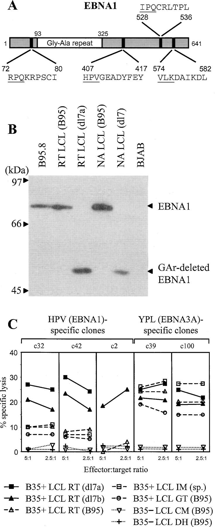
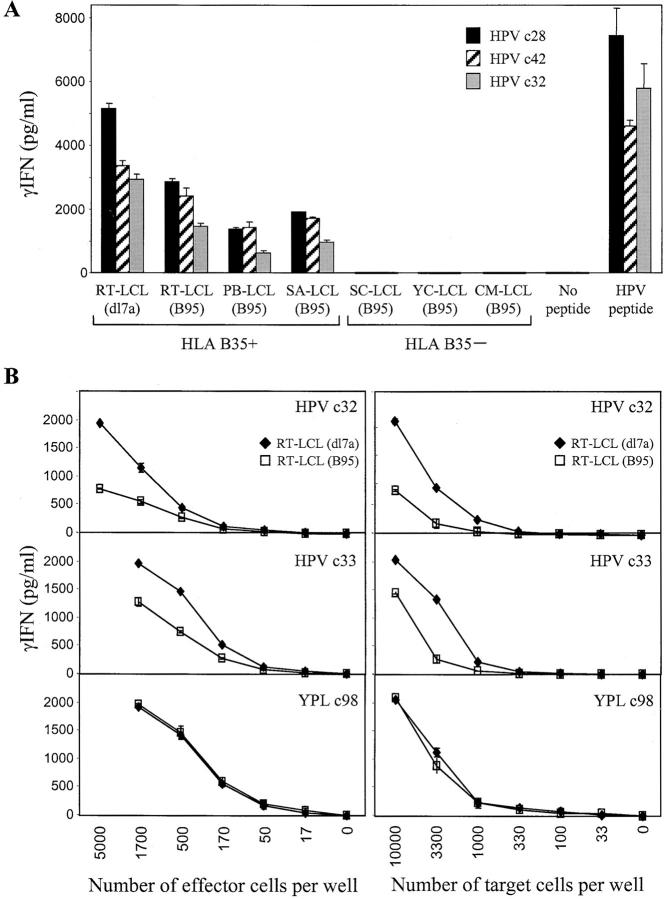
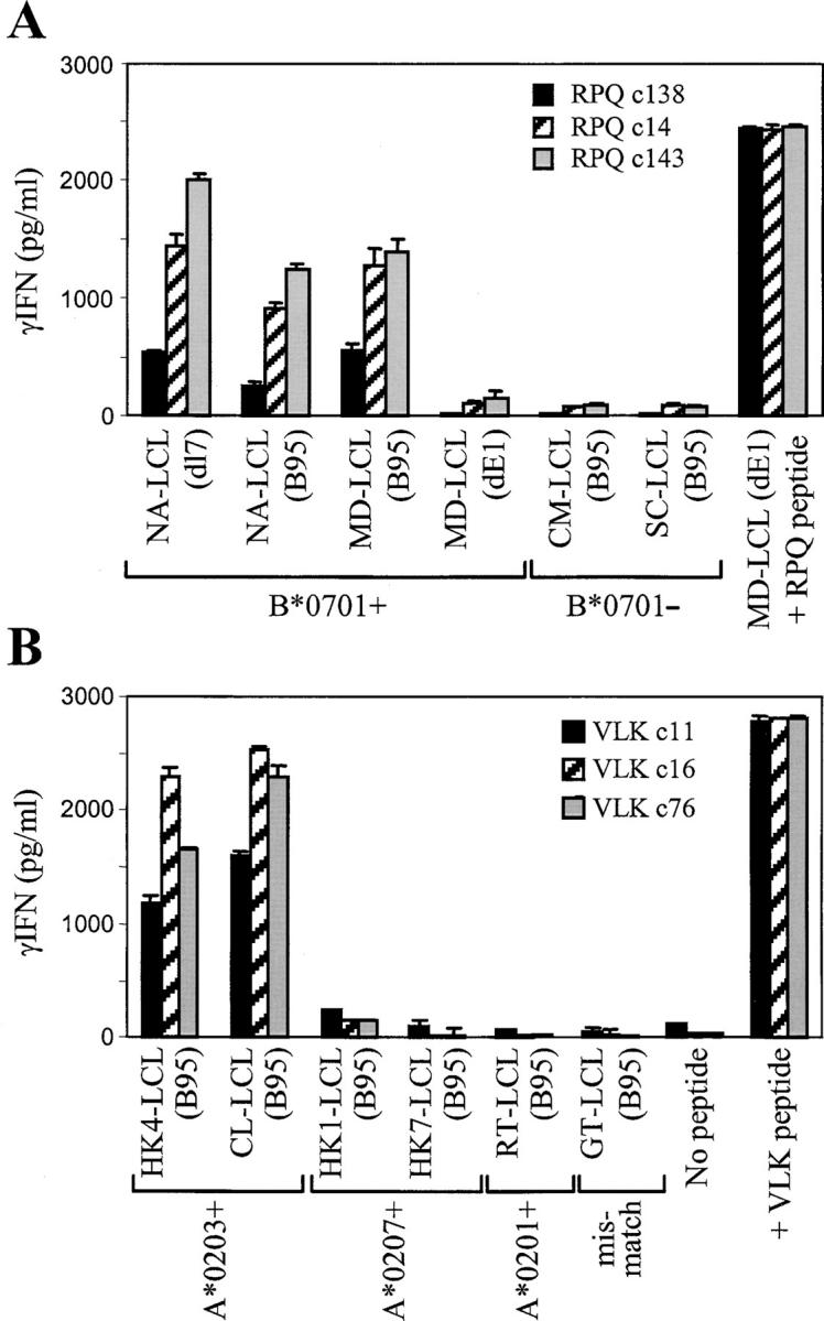
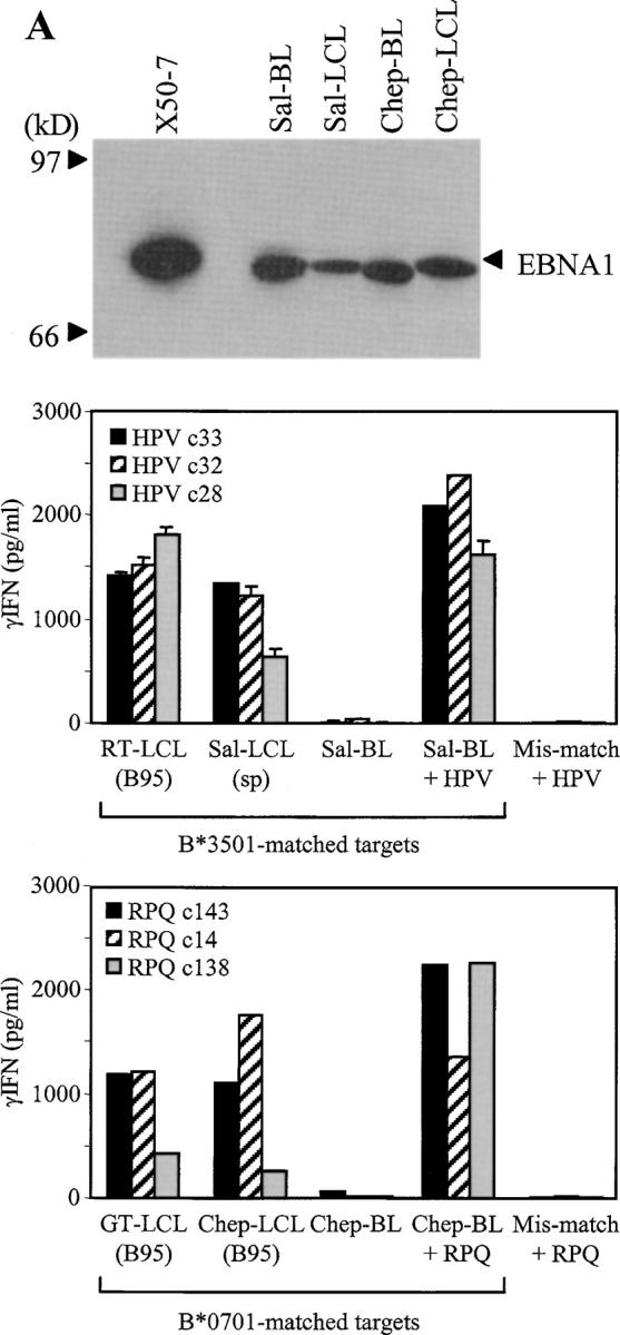
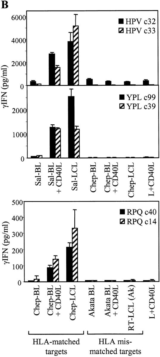
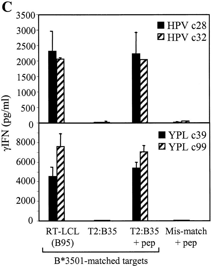
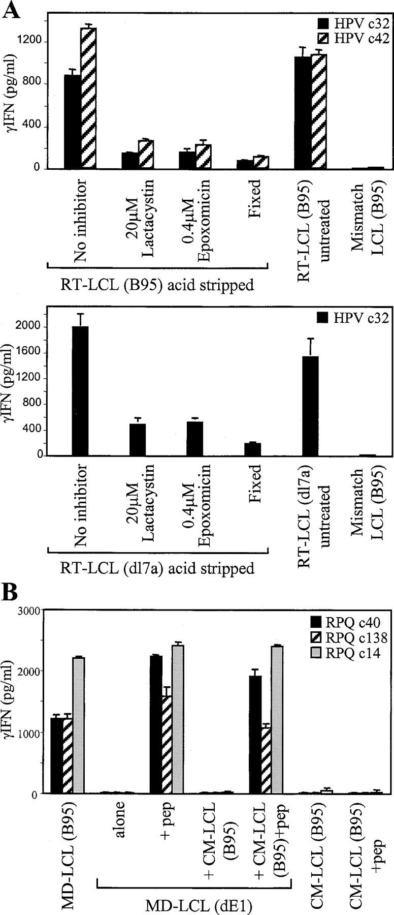
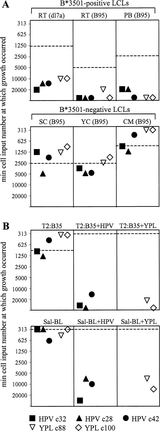
The Epstein-Barr virus (EBV) nuclear antigen (EBNA)1 contains a glycine-alanine repeat (GAr) domain that appears to protect the antigen from proteasomal breakdown and, as measured in cytotoxicity assays, from major histocompatibility complex (MHC) class I-restricted presentation to CD8+ T cells. This led to the concept of EBNA1 as an immunologically silent protein that although unique in being expressed in all EBV malignancies, could not be exploited as a CD8 target. Here, using CD8+ T cell clones to native EBNA1 epitopes upstream and downstream of the GAr domain and assaying recognition by interferon gamma release, we show that the EBNA1 naturally expressed in EBV-transformed lymphoblastoid cell lines (LCLs) is in fact presented to CD8+ T cells via a proteasome/peptide transporter-dependent pathway. Furthermore, LCL recognition by such CD8+ T cells, although slightly lower than seen with paired lines expressing a GAr-deleted EBNA1 protein, leads to strong and specific inhibition of LCL outgrowth in vitro. Endogenously expressed EBNA1 is therefore accessible to the MHC class I pathway despite GAr-mediated stabilization of the mature protein. We infer that EBNA1-specific CD8+ T cells do play a role in control of EBV infection in vivo and might be exploitable in the control of EBV+ malignancies.


Figures

Figure 1.
Figure 1.
EBNA1-specific CD8+ T cell recognition of LCLs measured by cytotoxicity. (A) Schematic diagram of wild-type EBNA1 protein showing the sequence of CD8+ epitopes. Numbers refer to coordinates in the 641–amino acid sequence of the B95.8 strain EBNA1 protein. (B) Immunoblot of SDS-PAGE–separated protein extracts from the B95.8 cell line, B95.8 virus-transformed LCLs, RT (B95), and NA (B95), expressing wild-type EBNA1 and from the corresponding dl7 virus transformants, RT (dl7a) and NA (dl7), expressing GAr-deleted EBNA1. BJAB is an EBV B lymphoma cell line. The blot is probed with the EBNA1-specific monoclonal antibody IH4. (C) Results of 5-h chromium release assays in which B*3501-restricted CD8+ T cell clones specific for the HPV (EBNA1) epitope or for the YPL (EBNA3A) epitope were used as effectors on a common panel of LCL targets. Targets included two dl7 virus transformants from B*3501+ donor RT, RT-LCL (dl7a), and RT-LCL (dl7b) expressing GAr-deleted EBNA1, three B*3501+ LCLs from donors RT, GT, and IM transformed either with B95.8 virus (for RT and GT) or by spontaneous outgrowth with the donor's own EBV strain (for IM), and two B95.8 LCLs from B*3501 donors CM and DH. Results are shown as percent-specific lysis at effector/target ratios of 5:1 and 2.5:1.
Figure 2.
Figure 2.
EBNA1-specific CD8+ T cell recognition of LCLs measured by IFN-γ release. (A) Three CD8+ T cell clones specific for the HPV/B*3501 epitope were tested against a panel of B*3501+ and B*3501 LCL targets transformed either with wild-type B95.8 virus or with the dl7 strain expressing GAr-deleted EBNA1. T cells and LCL targets were both seeded at 104 cells/well. As controls, T cells alone were tested in the presence of 10−7 M HPV epitope peptide or with no peptide. (B) The efficiency of RT-LCL (B95) and RT-LCL (dl7a) target recognition was assessed in titration assays using two HPV (EBNA1)–specific CD8+ T cell clones and one YPL (EBNA3A)–specific clone. The effect of titrating numbers of effector and target cells are shown on the left and right, respectively. When titrating numbers of effectors, LCL targets were added at 104 cells/well, and when titrating numbers of targets, effectors were added at 5 × 10³ cells/well. Results represent the mean values of IFN-γ release measured from triplicate cultures (+SD).
Figure 3.
Figure 3.
B*0701- and A*0203-restricted CD8+ T cell recognition of endogenously expressed EBNA1. (A) Three CD8+ T cell clones specific for the RPQ/B*0701 epitope were tested against a panel of B*0701+ and B*0701 LCL targets transformed with the wild-type EBV strain B95.8, against the B*0701+ NA-LCL (dl7) carrying an EBNA1 gene deleted for the GAr-coding sequence, or against the B*0701+ MD-LCL (dE1) carrying an EBV genome from which the EBNA1 gene has been completely deleted. As a positive control, T cells were tested against MD-LCL (dE1) cells that had been precoated with the target epitope peptide RPQ. (B) Three CD8+ T cell clones specific for the VLK/A*0203 epitope were tested against a panel of HLA-matched and -mismatched LCL targets transformed with the wild-type EBV strain B95.8. As a positive control, T cells were tested with 10−7 M VLK epitope peptide alone. T cells were tested at 104 cells/well and LCL targets were tested at 2 × 10⁴ cells/well. Results expressed as in Fig. 2.
Figure 4.
Figure 4.
Presentation of EBNA1 to CD8+ T cells does not occur in cells with defects in the proteasome/TAP-dependent processing pathway. (A) Top: EBNA1 immunoblot (as in Fig. 1 B) of protein extracts from the Sal-BL/Sal-LCL and Chep-BL/Chep-LCL pairings. X50-7 is a standard B95.8 virus-transformed LCL. Middle: Three CD8+ T cell clones specific for the HPV/B*3501 epitope were tested against the B*3501+ Sal-BL/Sal-LCL pair of targets and against Sal-BL cells precoated with HPV peptide. Control targets were the B*3501+ RT-LCL (B95) and an HLA-mismatched LCL precoated with HPV peptide. Bottom: Three T cell clones specific for the RPQ/B*0701 epitope were tested against the B*0701+ Chep-BL/Chep-LCL pair of targets and against Chep-BL cells precoated with RPQ peptide. Control targets were the B*0701+ GT-LCL (B95) and an HLA-mismatched LCL precoated with RPQ peptide. (B) Top panels: CD8+ T cell clones specific for the B*3501-restricted epitopes HPV (EBNA1) or YPL (EBNA3A) were tested against the Sal-BL/Sal-LCL pair of targets and against Sal-BL cells preexposed for 2 d to CD40 ligand–expressing mouse L cells (L+CD40L). Control targets included the Chep-BL/Chep-LCL pair, Chep-BL cells similarly exposed to L+CD40L cells, and L+CD40L cells themselves. Bottom: CD8+ T cell clones specific for the RPQ/B*0701 epitope were tested against the Chep-BL/Chep-LCL pair and against Chep-BL cells preexposed as described above to L+CD40L cells. Control targets included the HLA-mismatched Akata BL line, with and without preexposure to L+CD40L cells, the HLA-mismatched RT-LCL (Ak) transformed with the Akata virus strain, and L+CD40L cells themselves. (C) CD8+ T cell clones against the HPV and YPL epitopes were tested against the TAP T2:B35 LCL target line precoated or not with the relevant epitope peptide. Control targets included the B*3501+ RT-LCL (B95) and an HLA-mismatched LCL precoated with the relevant peptide. In all the above assays, T cells were tested at 1,000–10,000 cells per well and targets at 25,000 cells per well. Results expressed as in Fig. 2.
Figure 4.
Figure 4.
Presentation of EBNA1 to CD8+ T cells does not occur in cells with defects in the proteasome/TAP-dependent processing pathway. (A) Top: EBNA1 immunoblot (as in Fig. 1 B) of protein extracts from the Sal-BL/Sal-LCL and Chep-BL/Chep-LCL pairings. X50-7 is a standard B95.8 virus-transformed LCL. Middle: Three CD8+ T cell clones specific for the HPV/B*3501 epitope were tested against the B*3501+ Sal-BL/Sal-LCL pair of targets and against Sal-BL cells precoated with HPV peptide. Control targets were the B*3501+ RT-LCL (B95) and an HLA-mismatched LCL precoated with HPV peptide. Bottom: Three T cell clones specific for the RPQ/B*0701 epitope were tested against the B*0701+ Chep-BL/Chep-LCL pair of targets and against Chep-BL cells precoated with RPQ peptide. Control targets were the B*0701+ GT-LCL (B95) and an HLA-mismatched LCL precoated with RPQ peptide. (B) Top panels: CD8+ T cell clones specific for the B*3501-restricted epitopes HPV (EBNA1) or YPL (EBNA3A) were tested against the Sal-BL/Sal-LCL pair of targets and against Sal-BL cells preexposed for 2 d to CD40 ligand–expressing mouse L cells (L+CD40L). Control targets included the Chep-BL/Chep-LCL pair, Chep-BL cells similarly exposed to L+CD40L cells, and L+CD40L cells themselves. Bottom: CD8+ T cell clones specific for the RPQ/B*0701 epitope were tested against the Chep-BL/Chep-LCL pair and against Chep-BL cells preexposed as described above to L+CD40L cells. Control targets included the HLA-mismatched Akata BL line, with and without preexposure to L+CD40L cells, the HLA-mismatched RT-LCL (Ak) transformed with the Akata virus strain, and L+CD40L cells themselves. (C) CD8+ T cell clones against the HPV and YPL epitopes were tested against the TAP T2:B35 LCL target line precoated or not with the relevant epitope peptide. Control targets included the B*3501+ RT-LCL (B95) and an HLA-mismatched LCL precoated with the relevant peptide. In all the above assays, T cells were tested at 1,000–10,000 cells per well and targets at 25,000 cells per well. Results expressed as in Fig. 2.
Figure 5.
Figure 5.
Presentation of EBNA1 to CD8+ T cells is blocked by inhibitors of proteasome function and requires endogenously expressed antigen. (A) The B*3501+ RT-LCL (B95) and RT-LCL (dl7a) target cells were acid stripped to remove surface HLA class I–bound peptides, washed, and incubated either in standard medium or in the presence of 20 μM lactacystin or 0.4 μM epoxomicin. The target cells were then washed extensively and mixed with CD8+ T cell clones specific for the HPV/B*3501 epitopes. Control targets included cells of the same line that had been acid stripped and then immediately fixed with glutaraldehyde (Fixed), cells of the same line that had not been acid stripped or exposed to inhibitors (untreated), and cells of an HLA-mismatched B95.8 virus–transformed LCL. In a parallel experiment to check for nonspecific toxicity of the inhibitors, the above acid-stripped LCL cells were exposed to epitope peptide dilutions after their 2-h treatment with proteasome inhibitors, and then washed and tested as targets. These cells stimulated IFN-γ release after exposure to peptide concentrations as low as 10−10 M just as efficiently as did acid-stripped cells that had not been inhibitor treated. (B) The acceptor B*0701+/EBNA1 MD-LCL (dE1) was mixed with an equal number of cells from the donor B*0701/EBNA1+ CM-LCL (B95), and the mixture was cocultivated for 7 d before being used as targets for CD8+ T cell clones specific for the RPQ/B*0701 epitope. Control targets included the donor and acceptor LCLs alone, assayed pulsed or not pulsed with the RPQ peptide, the donor + acceptor cocultures pulsed with RPQ peptide, and the B*0701+ EBNA1+ cell line MD-LCL (B95). Results expressed as in Fig. 2.
Figure 6.
Figure 6.
EBNA1-specific CD8+ T cells inhibit the in vitro outgrowth of EBV-transformed LCLs of the appropriate HLA type. EBV+ target B cell lines were seeded in microtest plate wells at a range of input cell numbers (20,000–313 per well), either alone or in the presence of added CD8+ T cell clones (10,000 per well) specific for the HPV/B*3501 or YPL/B*3501 epitopes. For each target, results are expressed as the minimum target cell seeding required for successful outgrowth from the different types of coculture. The corresponding value for target cells in the absence of T cells is shown as a dotted horizontal line. Targets included B*3501+ and B*3501 LCLs transformed either with B95.8 virus or with the dl7 strain as well as the TAP LCL T2:B35 and the processing-deficient B*3501+ Sal-BL line, both either pulsed or not pulsed with the appropriate epitope peptide HPV or YPL. Cultures were assessed for cell growth after 4 wk. Control cultures set up with T cells alone contained no viable cells by that stage.

References

    1. Pamer, E., and P. Cresswell. 1998. Mechanisms of MHC class I–restricted antigen processing. Annu. Rev. Immunol. 16:323–358. - PubMed
    1. Koszinowski, U.H., and H. Hengel. 2002. Viral Proteins Counteracting Host Defences. Springer-Verlag, Berlin. 325 pp.
    1. Rickinson, A.B., and E. Kieff. 2001. Epstein-Barr virus. Fields Virology. D.M. Knipe, and P.M. Howley, editors. Lippincott Williams and Wilkins, Philadelphia. 2575–2627.
    1. Lee, M.A., M.E. Diamond, and J.L. Yates. 1999. Genetic evidence that EBNA-1 is needed for efficient, stable latent infection by Epstein-Barr virus. J. Virol. 73:2974–2982. - PMC - PubMed
    1. Levitskaya, J., M. Coram, V. Levitsky, S. Imreh, P.M. Steigerwald-Mullen, G. Klein, M.G. Kurilla, and M.G. Masucci. 1995. Inhibition of antigen processing by the internal repeat region of the Epstein-Barr virus nuclear antigen 1. Nature. 375:685–688. - PubMed

Publication types

MeSH terms