. 2004 May 17;32(9):2751-9.
doi: 10.1093/nar/gkh603. Print 2004.

CsdA, a cold-shock RNA helicase from Escherichia coli, is involved in the biogenesis of 50S ribosomal subunit

Affiliations

CsdA, a cold-shock RNA helicase from Escherichia coli, is involved in the biogenesis of 50S ribosomal subunit

Julie Charollais et al. Nucleic Acids Res. .

Abstract

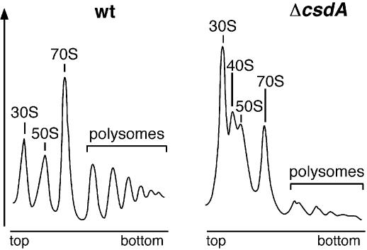
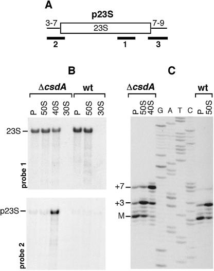
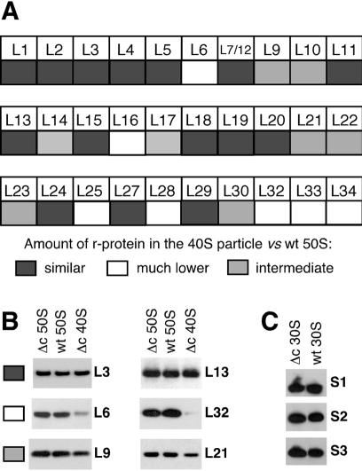
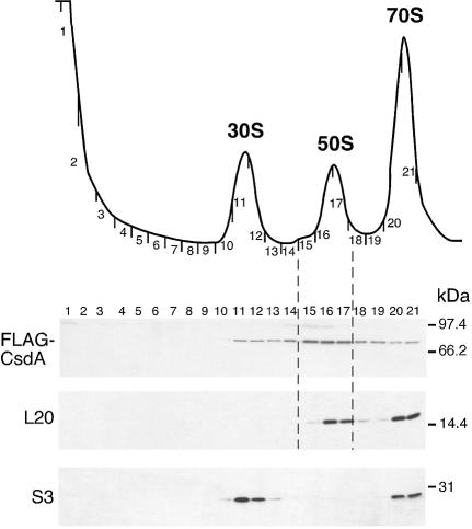
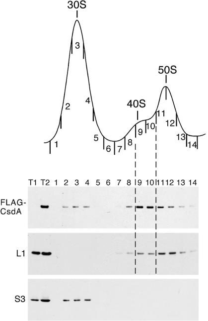
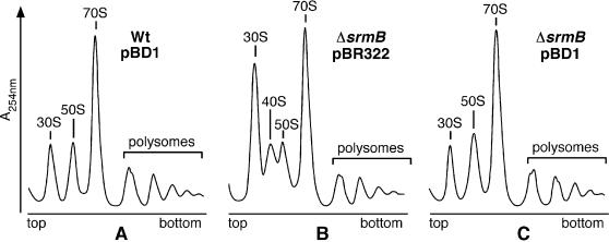
CsdA, a DEAD-box protein from Escherichia coli, has been proposed to participate in a variety of processes, such as translation initiation, gene regulation after cold-shock, mRNA decay and biogenesis of the small ribosomal subunit. Whether the protein really plays a direct role in these multiple processes is however, not clear. Here, we show that CsdA is involved in the biogenesis of the large rather than the small ribosomal subunit. Deletion of the csdA gene leads to a deficit in free 50S subunits at low temperatures and to the accumulation of a new particle sedimenting around 40S. Analysis of the RNA and protein contents of this particle indicates that it corresponds to a mis-assembled large subunit. Sucrose gradient fractionation shows that in wild-type cells CsdA associates mainly with a pre50S particle. Presumably the RNA helicase activity of CsdA permits a structural rearrangement during 50S biogenesis at low temperature. We showed previously that SrmB, another DEAD-box RNA helicase, is also involved in 50S assembly in E.coli. Our results suggest that CsdA is required at a later step than SrmB. However, over-expression of CsdA corrects the ribosome defect of the srmB-deleted strain, indicating that some functional overlap exists between the two proteins.


Figures

Figure 1
Figure 1
Deletion of csdA leads to a reduced growth at low temperature. wt and ΔcsdA strains transformed with either pBR322 or a derivative bearing the csdA gene (pBD1) were grown on tetracycline-LB plates at 37°C for 15 h or at 22°C for 2 days (at 22°C, the ΔcsdA/pBR322 colonies only become visible after longer incubation times).
Figure 2
Figure 2
Deletion of csdA leads to a deficiency in free 50S ribosomal subunits and to the accumulation of a new particle (40S). wt and ΔcsdA strains were grown in LB medium at 20°C. Polysome profiles were analysed as described in Materials and Methods. Ordinates refer to A254nm. Peaks corresponding to free 30S and 50S subunits, 70S ribosomes (free couples and monosomes), polysomes and 40S particles are indicated.
Figure 3
Figure 3
The ΔcsdA 40S particle contains a 23S rRNA precursor. Ribosomal particles from wt and ΔcsdA strains grown at 20°C were purified on two sucrose gradients. RNA extracted from these isolated particles was subjected to northern blot (B) and primer extension (C) analysis. (A) The p23S precursor results from RNase III cleavage of the initial 30S rrn transcript. Compared with mature 23S rRNA (open box), it caries 3 or 7 and 7–9 extra-nucleotides at its 5′ or 3′ ends, respectively [thin lines (26)]. The probes used for northern analysis are indicated by solid bars. (B) Equal amounts of RNA from 30S, 40S, 50S and polysome (P) fractions were separated on a 1% agarose gel, transferred to a nylon membrane and probed with the 5′-end-labeled oligonucleotides shown in (A). Upper panel, probe 1; lower panel, probe 2. Data obtained with probe 3 are not shown. The positions of precursor and mature 23S rRNA are indicated. (C) RNA from 40S, 50S and polysome (P) fractions was analysed by primer extension using a 33P-end-labelled primer. A sequencing ladder obtained with the same primer is shown (GATC). The 5′ end of mature 23S rRNA (M) and that of the p23S precursor (+3 and +7) are indicated.
Figure 4
Figure 4
r-Protein analysis of the 40S and 30S particles from ΔcsdA strain. The ΔcsdA and wt strains were grown at 20°C. r-Proteins were extracted from purified particles and identified and quantified by western blot as described in Materials and Methods. (A) Relative abundance of individual r-proteins in the 40S particle versus the 50S subunit. Grey squares, faint grey squares and white squares correspond to proteins present in similar, lower or much lower amount in the 40S particle, respectively. (B) Western blots illustrating the three classes of proteins defined in (A). Equivalent amounts of 40S particles or 50S subunits (see Materials and Methods) were analysed by western blotting using antibodies against the indicated r-proteins. Δc = ΔcsdA. (C) Western blots showing the presence of r-proteins S1–S3 in the 30S subunits from wt and ΔcsdA strains.
Figure 5
Figure 5
CsdA associates with ribosomes in wt cells at low temperature. (Top) Sedimentation profile showing the free subunits and 70S ribosomes from wt cells [BL21(DE3)-FLAG-csdA strain] grown at 16°C. (Bottom) Equal volumes of each fraction were subjected to western blotting using anti-FLAG, anti-L20 and anti-S3 antibodies.
Figure 6
Figure 6
CsdA associates with the 40S ribosomal particle from the ΔsrmB extract. (Top) Sedimentation profile showing the free subunits from ΔsrmB cells carrying a FLAG-tagged csdA gene (WJW45ΔsrmB FLAG-csdA). Cells were grown at 20°C. (Bottom) Equal volumes of each fraction were subjected to western blotting using anti-FLAG, anti-L1 and anti-S3 antibodies. Total proteins isolated from WJW45ΔsrmB (T1) and WJW45-FLAG-csdA (T2) strains were used as negative and positive controls for FLAG detection, respectively.
Figure 7
Figure 7
Over-expression of csdA suppresses the ΔsrmB ribosome defect. wt and ΔsrmB strains transformed with pBD1 (pBR322-csdA) or pBR322 plasmids were grown in tetracycline-LB medium at 20°C. Polysome profiles show the free ribosomal subunits, 70S ribosomes, polysomes and 40S particle.

References

    1. Linder P., Lasko,P.F., Ashburner,M., Leroy,P., Nielsen,P.J., Nishi,K., Schnier,J. and Slonimski,P.P. (1989) Birth of the DEAD box. Nature, 337, 121–122. - PubMed
    1. Tanner N.K. and Linder,P. (2001) DExD/H box RNA helicases: from generic motors to specific dissociation functions. Mol. Cell., 8, 251–262. - PubMed
    1. de la Cruz J., Kressler,D. and Linder,P. (1999) Unwinding RNA in Saccharomyces cerevisiae: DEAD-box proteins and related families. Trends Biochem. Sci., 24, 192–198. - PubMed
    1. Lorsch J.R. (2002) RNA chaperones exist and DEAD box proteins get a life. Cell, 109, 797–800. - PubMed
    1. Linder P., Tanner,N.K. and Banroques,J. (2001) From RNA helicases to RNPases. Trends Biochem. Sci., 26, 339–341. - PubMed

Publication types

MeSH terms