. 2004 May 25;101(21):7902-6.
doi: 10.1073/pnas.0402231101. Epub 2004 May 17.

Trigger factor binds to ribosome-signal-recognition particle (SRP) complexes and is excluded by binding of the SRP receptor

Affiliations

Trigger factor binds to ribosome-signal-recognition particle (SRP) complexes and is excluded by binding of the SRP receptor

Iwona Buskiewicz et al. Proc Natl Acad Sci U S A. .

Abstract

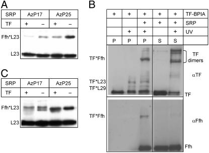
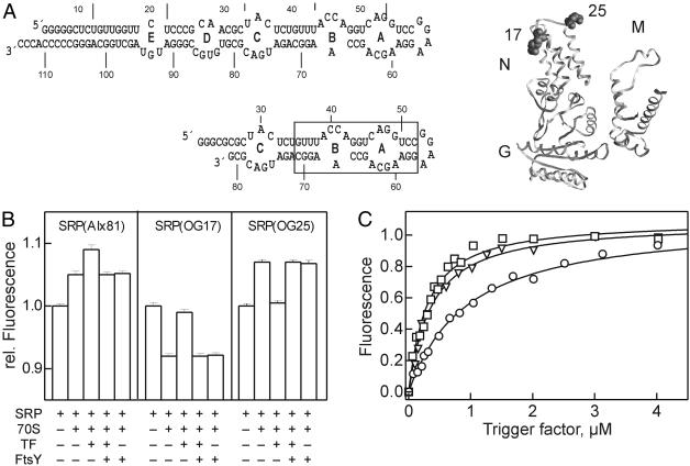
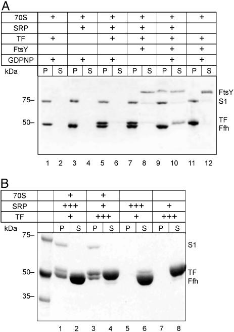
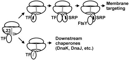
Trigger factor (TF) and signal recognition particle (SRP) bind to the bacterial ribosome and are both crosslinked to protein L23 at the peptide exit, where they interact with emerging nascent peptide chains. It is unclear whether TF and SRP exclude one another from their ribosomal binding site(s). Here we show that SRP and TF can bind simultaneously to ribosomes or ribosome nascent-chain complexes exposing a SRP-specific signal sequence. Based on changes of the crosslinking pattern and on results obtained by fluorescence measurements using fluorescence-labeled SRP, TF binding induces structural changes in the ribosome-SRP complex. Furthermore, we show that binding of the SRP receptor, FtsY, to ribosome-bound SRP excludes TF from the ribosome. These results suggest that TF and SRP sample nascent chains on the ribosome in a nonexclusive fashion. The decision for ribosome nascent-chain complexes exposing a signal sequence to enter SRP-dependent membrane targeting seems to be determined by the binding of SRP, which is stabilized by signal sequence recognition, and promoted by the exclusion of TF due to the binding of the SRP receptor to ribosome-bound SRP.


Figures

Fig. 1.
Fig. 1.
Ribosome binding of SRP and TF studied by crosslinking. (A) Crosslinking of SRP(AzP17/25) to vacant ribosomes. Ribosomes (1 μM), SRP(AzP17/25) (1 μM), and TF (10 μM) were irradiated with UV light, samples were centrifuged, and ribosomal pellets were analyzed by SDS gel electrophoresis and immunoblotting using antibodies against ribosomal protein L23 (10). Crosslinked proteins are indicated by asterisks, e.g., Ffh*L23. (B) Crosslinking of TF–BPIA to vacant ribosomes. Ribosomes (2 μM), SRP (10 μM), and TF–BPIA (10 μM) were mixed and irradiated with UV light; after ultracentrifugation, ribosomal pellets (P) and supernatants (S) were analyzed by SDS gel electrophoresis and immunoblotting (9). For staining, antibodies against TF (αTF) and Ffh (αFfh) were used. (C) Crosslinking from SRP to lep-RNC. UV-induced crosslinking was performed with SRP(AzP17/25) (10 nM) and lep-RNC (10 nM) (Materials and Methods) in the absence and presence of TF (1 μM), and samples were analyzed as in A.
Fig. 3.
Fig. 3.
Ribosome binding of SRP, TF, and FtsY monitored by fluorescence. (A)(Left) Two-dimensional structure models of 4.5S RNA and 4.5S RNA(21–81). The approximate binding site of Ffh is boxed. (Right) Positions of OG labels in the N domain of Ffh. The arrangement of NG and M domains of Ffh is based on fluorescence data (I.B., unpublished data). (B) TF/FtsY binding to ribosome–SRP(Alx81) or ribosome–SRP(OG17/25) complexes. Fluorescence measurements were performed in buffer A containing 1 mM GDPNP at 20°C; excitation/emission was at 535/565 nm (Alx) or 470/517 nm (OG). To fluorescent SRP (0.1 μM) were added 70S ribosomes (0.1 μM), TF (1.2 μM), and FtsY (0.1 μM); the order of addition of TF and FtsY had no influence (data not shown). (C) TF titrations of ribosome–SRP(Alx81) complexes (0.1 μM) with TF (squares), TF (1–144) (triangles), or TF(FRK/AAA) (circles). Titrations were performed in buffer A at 20°C in the presence of 1 mM GDPNP. Data were evaluated by nonlinear fitting on the basis of 1:1 stoichiometric binding (20, 22); the following Kd values were obtained: TF, 0.33 ± 0.03 μM; TF(1–144), 0.30 ± 0.03 μM; TF(FRK/AAA), 1.0 ± 0.05 μM.
Fig. 2.
Fig. 2.
Ribosome binding of SRP, TF, and FtsY examined by ultracentrifugation. (A) Equimolar concentrations of ribosomes, SRP, TF, and FtsY. Components (1 μM each) were mixed in various combinations in buffer A (50 mM Tris·HCl, pH 7.5/70 mM NH4Cl/30 mM KCl/7 mM MgCl2), containing 1 mM GDPNP where indicated, and incubated for 10 min at 37°C. Samples (100 μl) were centrifuged at 436,000 × g (Sorvall M120GX) for 30 min at 4°C. Ribosomal pellets (P) and supernatants (S) were analyzed by SDS gel electrophoresis, and proteins were stained with Coomassie blue. S1, ribosomal protein S1 (69 kDa); the other ribosomal proteins migrated out of the gel. (B) Excess of SRP or TF over ribosomes. The experiment was performed as in A with ribosomes (1 μM, lanes 1–4), SRP (1 μM, lanes 3–6; 30 μM, lanes 1 and 2), and TF (1 μM, lanes 1, 2, 5, and 6; 30 μM, lanes 3, 4, 7, and 8); controls without ribosomes are shown in lanes 5–8. GDPNP (1 mM) was present in all samples.
Fig. 4.
Fig. 4.
Interplay of TF, SRP, and FtsY on the ribosome. See text for the description of steps. The nascent peptide (gray line) is depicted as running through the exit tunnel of the large ribosomal subunit; the SRP-specific signal sequence is depicted as a black box. The peptide exit region of the ribosome is depicted in a highly simplified manner, showing only protein L23, which forms part of the attachment sites of both TF and SRP.

Similar articles

Cited by

References

    1. Ban, N., Nissen, P., Hansen, J., Moore, P. B. & Steitz, T. A. (2000) Science 289, 905–920. - PubMed
    1. Harms, J., Schluenzen, F., Zarivach, R., Bashan, A., Gat, S., Agmon, I., Bartels, H., Franceschi, F. & Yonath, A. (2001) Cell 107, 679–688. - PubMed
    1. Frydman, J. (2001) Annu. Rev. Biochem. 70, 603–647. - PubMed
    1. Hartl, F. U. & Hayer-Hartl, M. (2002) Science 295, 1852–1858. - PubMed
    1. Valent, Q. A., Kendall, D. A., High, S., Kusters, R., Oudega, B. & Luirink, J. (1995) EMBO J. 14, 5494–5505. - PMC - PubMed

Publication types

MeSH terms

LinkOut - more resources