. 2004 May 25;101(21):7931-6.
doi: 10.1073/pnas.0400973101. Epub 2004 May 17.

The crystal structure of xanthine oxidoreductase during catalysis: implications for reaction mechanism and enzyme inhibition

Affiliations

The crystal structure of xanthine oxidoreductase during catalysis: implications for reaction mechanism and enzyme inhibition

Ken Okamoto et al. Proc Natl Acad Sci U S A. .

Abstract

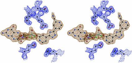
Molybdenum is widely distributed in biology and is usually found as a mononuclear metal center in the active sites of many enzymes catalyzing oxygen atom transfer. The molybdenum hydroxylases are distinct from other biological systems catalyzing hydroxylation reactions in that the oxygen atom incorporated into the product is derived from water rather than molecular oxygen. Here, we present the crystal structure of the key intermediate in the hydroxylation reaction of xanthine oxidoreductase with a slow substrate, in which the carbon-oxygen bond of the product is formed, yet the product remains complexed to the molybdenum. This intermediate displays a stable broad charge-transfer band at approximately 640 nm. The crystal structure of the complex indicates that the catalytically labile Mo-OH oxygen has formed a bond with a carbon atom of the substrate. In addition, the MoS group of the oxidized enzyme has become protonated to afford Mo-SH on reduction of the molybdenum center. In contrast to previous assignments, we find this last ligand at an equatorial position in the square-pyramidal metal coordination sphere, not the apical position. A water molecule usually seen in the active site of the enzyme is absent in the present structure, which probably accounts for the stability of this intermediate toward ligand displacement by hydroxide.


Figures

Fig. 1.
Fig. 1.
Structures of molybdopterin cofactor (a), FYX-051 (b), and 2-OH-FYX-051 (c). (d) Time-dependent inhibition of XO by FYX-051. Urate formation was followed by the absorbance change at 295 nm in 0.1 M pyrophosphate buffer (pH 8.5) containing 0.15 mM xanthine and 0.2 mM EDTA in the presence (solid) or absence (dashed) of 30 nM FYX-051 under aerobic conditions at 25°C. Reactions were started by adding 1 nM (final concentration) of fully active XO.
Fig. 4.
Fig. 4.
Stereo representation of the structure in the active site of XDH with bound FYX-051. FYX-051 (magenta), molybdopterin (cyan), and catalytically important amino acid residues (CPK-atom colored) are illustrated as stick models on a ribbon model background.
Fig. 5.
Fig. 5.
Stereo representation of the FoFc electron density corresponding to molybdopterin-FYX-051 (brown), contoured at a 3.0 σ cutoff, and the 2FoFc electron density corresponding to selected amino acid residues (blue) in the active site of the enzyme, contoured at a 1.5 σ cutoff. The FoFc density was calculated before introducing the Mo-cofactor and FYX-051 molecules into the crystallographic model to avoid model bias.
Fig. 6.
Fig. 6.
Electron density map around the molybdenum atom. The density was calculated before introducing the Mo-cofactor and inhibitor molecule FYX-051 into the crystallographic refinement. (a) FoFc electron density map, contoured at a 3.0 σ cutoff, surrounding the molybdenum ion and its immediate ligands in the complex. (b) Same view as in a but at a 15.0 σ cutoff with significant electron density still present at the positions assigned to sulfur atoms but absent at the position of the oxygen atom. (c) FoFc electron density map, contoured at a 3.0 σ cutoff, between the molybdenum ion and C2 atom of FYX-051–XDH. (d) FoFc electron density map of the view from the apical position of the molybdopterin-FYX-051 complex contoured at a 3.0 σ cutoff. (e) Same view as in d but at a 15.0 σ cutoff with significant electron density still present at the positions assigned to sulfur atoms but absent at the position of the oxygen atom.
Fig. 7.
Fig. 7.
Proposed mechanism initiated by base-assisted nucleophilic attack of Mo—OH on heterocycles, with subsequent hydride transfer to produce the reaction intermediate (c) whose structure has been analyzed in this report. The subsequent oxidation occurs via d or/and e with varying ratio depending on the substrate used.
Fig. 2.
Fig. 2.
(a) The absorption spectra of XOR after mixing with FXY-051 under anaerobic conditions. The spectrum of the enzyme (≈100% active) before mixing the enzyme with FXY-051 (black solid line), immediately after (dotted), and 30 min after exposure to air (red). (Inset) Difference spectrum between black solid and red lines. (b) The absorption spectra of dithionite-reduced XOR mixed with 2-OH-FYX-051 under anaerobic conditions. Original oxidized enzyme (black solid) dithionite reduced enzyme (open circle) enzyme after mixing with 2-OH-FYX-051(closed circle) and after exposure to air (red). (Inset) Difference spectrum between black solid and red lines.
Fig. 3.
Fig. 3.
(a) HPLC analysis of the compound released from the XDH-FYX-051 complex after protein denaturation. (Upper) Authentic FYX-051 and 2-OH-FYX-051. (Lower) The released compound. (b) Liquid chromatography–MS analysis of the compound released from the XDH-FYX-051 complex after protein denaturation. (Top) Authentic FYX-051; (Middle) 2-OH-FYX-051; (Bottom) the released compound.

Similar articles

Cited by

References

    1. Hille, R. & Nishino, T. (1995) FASEB J. 9, 995–1003. - PubMed
    1. Hille, R. (1996) Chem. Rev. 96, 2757–2816. - PubMed
    1. Elion, G. B. (1989) Science 244, 41–47. - PubMed
    1. Massey, V. & Harris, C. M. (1997) Biochem. Soc. Trans. 25, 750–755. - PubMed
    1. Enroth, C., Eger, B. T., Okamoto, K., Nishino, T., Nishino, T. & Pai, E. F. (2000) Proc. Natl. Acad. Sci. USA 97, 10723–10738. - PMC - PubMed

Publication types

MeSH terms

Associated data

LinkOut - more resources