. 2004 May 25;101(21):7891-6.
doi: 10.1073/pnas.0401167101. Epub 2004 May 17.

The mechanism of transmembrane S-nitrosothiol transport

Affiliations

The mechanism of transmembrane S-nitrosothiol transport

Yanhong Zhang et al. Proc Natl Acad Sci U S A. .

Abstract

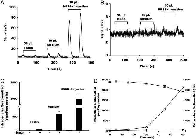
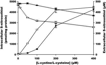
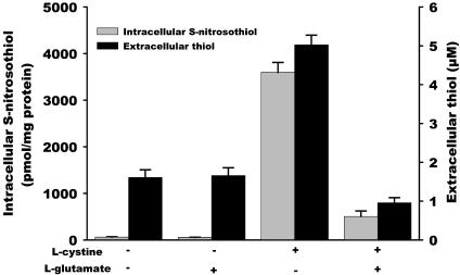
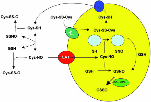
S-nitrosothiols have been suggested to play an important role in nitric oxide (NO)-mediated biological events. However, the mechanisms by which an S-nitrosothiol (or the S-nitroso functional group) is transferred across cell membrane are still poorly understood. We have demonstrated previously that the degradation of S-nitrosoglutathione (GSNO) by cells absolutely required the presence of cystine in the extracellular medium and proposed a mechanism that involved the reduction of cystine to cysteine, followed by the reaction of cysteine with GSNO to form S-nitrosocysteine (CysNO), mixed disulfides, and nitrosyl anion. In the present study we have assessed the effect of cystine on the transfer of the S-nitroso functional group from the extracellular to the intracellular space. Using RAW 264.7 cells, we found that the presence of L-cystine enhanced GSNO-dependent S-nitrosothiol uptake, increasing the intracellular S-nitrosothiol level from approximately 60 pmol/mg of protein to approximately 3 nmol/mg of protein. The uptake seems to depend on the reduction of L-cystine to L-cysteine, which involves the xc- amino acid transport system, the transnitrosation between GSNO and L-cysteine to form L-CysNO, and uptake of L-CysNO via amino acid transport system L. Compared with GSNO, (Z)-1-[N-(3-ammoniopropyl)-N-[4-(3-aminopropylammonio)butyl]-amino]diazen-1-ium-1,2-diolate, an NO donor, is much less effective at intracellular S-nitrosothiol formation in the presence of L-cystine or L-cysteine, suggesting that the biochemical changes that occur after exposure of cells to S-nitrosothiol, with respect to thiol chemistry, are distinctly different from those observed with NO.


Figures

Fig. 1.
Fig. 1.
The effect of l-cystine on intracellular S-nitrosothiol formation. RAW 264.7 cells were treated with GSNO (500 μM) for 60 min in HBSS, FBS-free medium, or HBSS supplemented with l-cystine (200 μM). Representative chemiluminescence traces of sulfanilamide-treated (A) or HgCl2/sulfanilamide-treated (B) samples are shown. (C) Quantification of S-nitrosothiol content in the cell lysate. Data represent mean ± SEM. (D) Time course of the formation of intracellular S-nitrosothiol in the presence (○) or absence (•) of l-cystine as well as the decay of extracellular S-nitrosothiol level in the presence of l-cystine (▴). Data represent mean ± SEM (n = 3).
Fig. 2.
Fig. 2.
Concentration response of l-cystine- or l-cysteine-mediated intracellular S-nitrosothiol formation and extracellular S-nitrosothiol decay. RAW 264.7 cells were treated with GSNO (500 μM) in HBSS for 60 min in the presence of various concentrations of l-cystine (filled symbols) or l-cysteine (open symbols). The S-nitrosothiol content in the cell lysate (• and ○) was measured by chemiluminescence. The S-nitrosothiol level in HBSS (▴ and ▵) was detected by UV-visible spectroscopy. Data represent mean ± SEM (n = 3).
Fig. 3.
Fig. 3.
The effect of l-glutamate on the formation of intracellular S-nitrosothiol and extracellular thiol. S-nitrosothiol formation (gray bars): RAW 264.7 cells were treated with GSNO (500 μM) for 60 min in the absence or presence of l-cystine (200 μM) and l-glutamate (3 mM). The S-nitrosothiol content in the cell lysate was measured by chemiluminescence. Thiol formation (black bars): RAW 264.7 cells were incubated in HBSS for 60 min in the absence and presence of l-cystine and l-glutamate. The thiol level in the HBSS was detected by a DTNB assay. Data represent mean ± SEM (n = 3).
Scheme 1.
Scheme 1.
A model for l-cystine-mediated S-nitrosothiol uptake.

References

    1. Ramachandran, N., Root, P., Jiang, X.-M., Hogg, P. J. & Mutus, B. (2001) Proc. Natl. Acad. Sci. USA 98, 9539–9544. - PMC - PubMed
    1. Zai, A., Rudd, M. A., Scribner, A. W. & Loscalzo, J. (1999) J. Clin. Invest. 103, 393–399. - PMC - PubMed
    1. De Groote, M. A., Granger, D., Xu, Y., Campbell, G., Prince, R. & Fang, F. C. (1995) Proc. Natl. Acad. Sci. USA 92, 6399–6403. - PMC - PubMed
    1. Hogg, N., Singh, R. J., Konorev, E., Joseph, J. & Kalyanaraman, B. (1997) Biochem. J. 323, 477–481. - PMC - PubMed
    1. Pawloski, J. R., Hess, D. T. & Stamler, J. S. (2001) Nature 409, 622–626. - PubMed

Publication types

LinkOut - more resources