. 2004 Jun 1;101(22):8461-6.
doi: 10.1073/pnas.0401440101. Epub 2004 May 17.

Functional photoreceptor loss revealed with adaptive optics: an alternate cause of color blindness

Affiliations

Functional photoreceptor loss revealed with adaptive optics: an alternate cause of color blindness

Joseph Carroll et al. Proc Natl Acad Sci U S A. .

Abstract

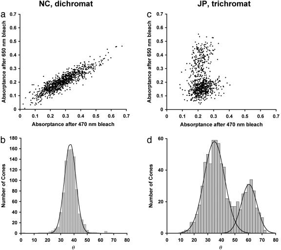
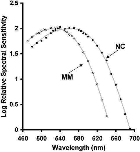
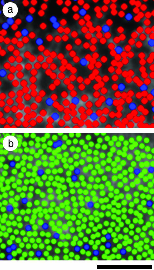
There is enormous variation in the X-linked L/M (long/middle wavelength sensitive) gene array underlying "normal" color vision in humans. This variability has been shown to underlie individual variation in color matching behavior. Recently, red-green color blindness has also been shown to be associated with distinctly different genotypes. This has opened the possibility that there may be important phenotypic differences within classically defined groups of color blind individuals. Here, adaptive optics retinal imaging has revealed a mechanism for producing dichromatic color vision in which the expression of a mutant cone photopigment gene leads to the loss of the entire corresponding class of cone photoreceptor cells. Previously, the theory that common forms of inherited color blindness could be caused by the loss of photoreceptor cells had been discounted. We confirm that remarkably, this loss of one-third of the cones does not impair any aspect of vision other than color.


Figures

Fig. 1.
Fig. 1.
Retinal images from the right eyes of a trichromat and two dichromats. JP (age 28) is a trichromat, MM (age 32) is a protanope, and NC (age 26) is a deuteranope. Images are at ≈1° eccentricity from nasal (a, c, and e) or temporal (b, d, and f) retina. MM (c and d) was classified as a protanope, and NC (e and f) was classified as a deuteranope based on Rayleigh match data and performance on standard color vision tests. Images from trichromat JP (a and b) are shown for comparison. (Scale bar, 20 μm.)
Fig. 2.
Fig. 2.
Scatter plots and histograms of individual cone absorptances. (a) Scatter plot shows individual cone absorptances from NC's retinal images after the 470- and 650-nm selective bleaches. Cone absorptance was taken as the average value computed within a 0.4-arcmin square region centered on the cone. A total of 932 non-S cones in a 0.05-mm2 area were analyzed (eccentricity of 0.5° temporal retina). (b) Histogram of individual cone absorptances, where the number of cones is plotted as a function of angle in the scatter plot in a. Solid line represents the best-fitting Gaussian curve (r2 = 0.99). The residual single mode is indicative of a single L/M cone type (17). (c) Same as in a, but for JP (trichromat). A total of 741 non-S cones in a 0.03-mm2 area were analyzed (eccentricity of 1° temporal retina). L cones absorb relatively less after the 650-nm bleach and relatively more after the 470-nm bleach than the M cones do, thus they appear closer to the abscissa. (d) The histogram for JP. A sum of two Gaussian curves was fit to the histogram (solid lines, r2 = 0.97). The estimated L:M ratio from this analysis is 2.4:1.
Fig. 3.
Fig. 3.
Relative spectral sensitivity functions for MM and NC. Spectral sensitivity was measured with the flicker photometric ERG. Filled circles are from the right eye of NC, and open circles are from the right eye of MM. Solid lines represent vitamin-A1 visual pigment templates with λmax values of 559 nm and 527 nm, consistent with typical deuteranope and protanope values, respectively (see text).
Fig. 4.
Fig. 4.
Pseudocolor image of the dichromatic cone mosaic. Blue, green, and red colors represent the S, M, and L cones, respectively. (a) Subject NC's inferior retina at an eccentricity of 0.75°. All cones are of the S or L type (see text for details). (b) Subject MM's nasal retina at an eccentricity of 1°. All cones are of the S or M type, as indicated from previous results (17) as well as data presented here (see Fig. 3). (Scale bar, 50 μm.)

References

    1. Berendschot, T. T., van de Kraats, J. & van Norren, D. (1996) J. Physiol. 492, 307-314. - PMC - PubMed
    1. Cicerone, C. M. & Nerger, J. L. (1989) Vision Res. 29, 1587-1595. - PubMed
    1. Wesner, M. F., Pokorny, J., Shevell, S. K. & Smith, V. C. (1991) Vision Res. 31, 1021-1037. - PubMed
    1. Kremers, J., Usui, T., Scholl, H. P. N. & Sharpe, L. T. (1999) Invest. Ophthalmol. Vis. Sci. 40, 920-930. - PubMed
    1. Nathans, J., Thomas, D. & Hogness, D. S. (1986) Science 232, 193-202. - PubMed

Publication types

Substances