. 2004 May 25;101(21):7988-93.
doi: 10.1073/pnas.0401897101. Epub 2004 May 17.

Cytomegalovirus cell death suppressor vMIA blocks Bax- but not Bak-mediated apoptosis by binding and sequestering Bax at mitochondria

Affiliations

Cytomegalovirus cell death suppressor vMIA blocks Bax- but not Bak-mediated apoptosis by binding and sequestering Bax at mitochondria

Damien Arnoult et al. Proc Natl Acad Sci U S A. .

Abstract

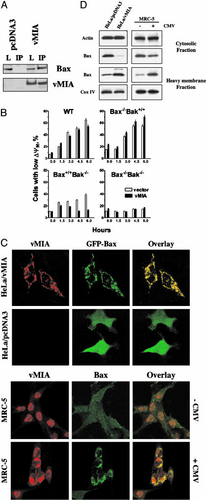
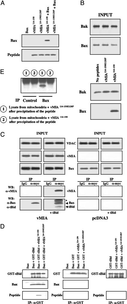
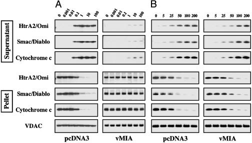
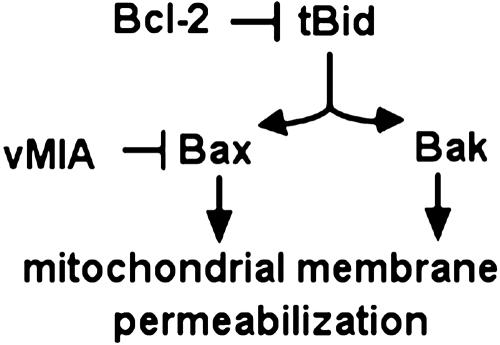
We report that the cytomegalovirus-encoded cell death suppressor vMIA binds Bax and prevents Bax-mediated mitochondrial membrane permeabilization by sequestering Bax at mitochondria in the form of a vMIA-Bax complex. vMIA mutants with a defective mitochondria-targeting domain retain their Bax-binding function but not their ability to suppress mitochondrial membrane permeabilization or cell death. vMIA does not seem to either specifically associate with Bak or suppress Bak-mediated mitochondrial membrane permeabilization. Recent evidence suggests that the contribution of Bax and Bak in the mitochondrial apoptotic signaling pathway depends on the distinct phenotypes of cells, and it appears from our data that vMIA is capable of suppressing apoptosis in cells in which this pathway is dominated by Bax, but not in cells where Bak also plays a role.


Figures

Fig. 1.
Fig. 1.
(A) Bax is associated with vMIA in cells expressing vMIA. Lysates (L) from HeLa cells lysed in a CHAPS-containing buffer (LBC, see Experimental Procedures) constitutively expressing myc-tagged vMIA, or from control cells (pcDNA3 transfected) were immunoprecipitated (IP) with anti-myc beads, bound proteins were separated by SDS/PAGE and examined by Western blotting with either anti-Bax or anti-myc. Equal amounts of HeLa/pcDNA3 and HeLa/vMIA lysates (by total protein) were loaded. The “inputs” represent 10% of the starting materials for IP. (B) vMIA protects Bax-expressing MEFs that lack Bak, but fails to protect Bak-expressing MEFs from staurosporine-induced apoptosis. The loss of the mitochondrial membrane potential ΔΨM was used as an index for apoptosis (see text). The data presented as the means ± SD (three independent experiments). (C) GFP–Bax colocalizes with vMIA in transiently transfected cells. Cells stably transfected with vector only (HeLa/pcDNA3) or myc-tagged vMIA (HeLa/vMIA) were transfected with EGFP–Bax. Eighteen hours after transfection, cells were stained with anti-myc recognizing the myc-tag of vMIA (red). Colocalization of green and red fluorescence is indicated by a yellow blend. Endogenous Bax colocalizes with vMIA in human fibroblasts infected with human CMV (TownevarRIT, 48 h after infection). Cells were stained with rabbit anti-vMIA antiserum (green) and mouse monoclonal anti-Bax (red). Colocalization of green and red fluorescence is indicated by a yellow blend. (D) vMIA induces relocation of endogenous Bax to mitochondria. Cytosolic and heavy membrane fractions of HeLa/pcDNA3 and HeLa/vMIA cells and of MRC-5 human fibroblasts infected or not with human CMV (TownevarRIT, 72 h after infection) were analyzed by Western blotting for the presence of Bax. As control for loading, actin was used in the cytosolic fraction and Cox IV in the heavy membrane fraction. Visual observation of the infected populations of fibroblasts indicated that a fraction of cells remained noninfected, consistent with some remaining cytoplasmic Bax.
Fig. 2.
Fig. 2.
(A) A synthetic peptide vMIA114-150 has specific affinity for Bax. Biotinylated vMIA114-150 (1 μM) and control biotinylated vMIA114-150E128P (1μM), containing a point mutation that inactivates cell-death-suppressing activity of vMIA were incubated with either recombinant Bax (100 nM) or buffer alone, pulled down with streptavidin-agarose, separated by SDS/PAGE, and examined by Western blotting with horseradish peroxidase–streptavidin and anti-Bax. (B) vMIA114-150 binds endogenous Bax, but not endogenous Bak. Isolated mitochondria (500 μg) were incubated with biotinylated vMIA114-150 (10 μg), control biotinylated vMIA114-150E128P (10 μg), or buffer alone, pulled down with streptavidin-agarose, separated by SDS/PAGE, and analyzed by Western blotting. As a control, “input” represents 2% of the starting material. (C) vMIA does not prevent the binding of tBid to Bax. Isolated mitochondria from HeLa/vMIA (vMIA mitochondria) (500 μg) or from HeLa/pcDNA3 (500 μg) were incubated with tBid (100 nM) or buffer alone, then vMIA was immunoprecipitated (IP) with anti-myc by using an irrelevant antibody as a control, the samples were separated by SDS/PAGE, and Bax, tBid, and vMIA were then detected by Western blotting (WB). Asterisk indicates a band that may be either of a nonspecific nature, or a covalently modified form of Bax or tBid. As a control, “input” represents 2% of the starting material. (D) Recombinant Bax (100 nM) was incubated with GST–tBid (100 nM) in the presence or absence of a 10-fold molar excess of vMIA114-150 or vMIA114-150 E128P (each at 1 μM), then GST–tBid was immunoprecipitated (IP) with an anti-GST, and bound proteins were separated by SDS/PAGE and detected by Western blotting (Left). In control samples, Bax (100 nM) was incubated with GST (100 nM) in the presence or absence of the biotinylated peptides (1 μM), and GST–tBid was incubated with the biotinylated peptides (1 μM) to see whether they bind GST–tBid (Right). (E) Mitochondria isolated from HeLa/pcDNA3 were incubated either with vMIA114-150 (1 μM) or vMIA114-150E128P (1 μM) as a control. Peptides were pulled down as in B, then Bax was immunoprecipitated (IP) from the remaining supernatant of the mitochondrial lysate, and the immunoprecipitate was examined for the presence of Bax. An irrelevant antibody was used as a control.
Fig. 3.
Fig. 3.
(A and B) vMIA prevents mitochondrial membrane permeabilization induced by recombinant tBid (concentrations are shown on top in nM) (A), but not by recombinant oligomeric Bax (concentrations are shown on top in nM) (B), see text for explanations. Equal loading of the mitochondrial pellet was verified by using VDAC.
Fig. 4.
Fig. 4.
A schematic representation of suppression of the tBid-mediated apoptotic pathway by vMIA.

References

    1. Goldmacher, V. S., Bartle, L. M., Skaletskaya, A., Dionne, C. A., Kedersha, N. L., Vater, C. A., Han, J. W., Lutz, R. J., Watanabe, S., Cahir McFarland, E. D., et al. (1999) Proc. Natl. Acad. Sci. USA 96, 12536–12541. - PMC - PubMed
    1. McCormick, A. L., Skaletskaya, A., Barry, P. A., Mocarski, E. S. & Goldmacher, V. S. (2003) Virology 316, 221–233. - PubMed
    1. Goldmacher, V. S. (2002) Biochimie 84, 177–185. - PubMed
    1. Yang, J., Liu, X., Bhalla, K., Kim, C. N., Ibrado, A. M., Cai, J., Peng, T. I., Jones, D. P. & Wang, X. (1997) Science 275, 1129–1132. - PubMed
    1. Kluck, R. M., Bossy-Wetzel, E., Green, D. R. & Newmeyer, D. D. (1997) Science 275, 1132–1136. - PubMed

MeSH terms