. 2004 May 18:2:7.
doi: 10.1186/1741-7007-2-7.

PI3 kinase is important for Ras, MEK and Erk activation of Epo-stimulated human erythroid progenitors

Affiliations

PI3 kinase is important for Ras, MEK and Erk activation of Epo-stimulated human erythroid progenitors

Enrico K Schmidt et al. BMC Biol. .

Abstract

Background: Erythropoietin is a multifunctional cytokine which regulates the number of erythrocytes circulating in mammalian blood. This is crucial in order to maintain an appropriate oxygen supply throughout the body. Stimulation of primary human erythroid progenitors (PEPs) with erythropoietin (Epo) leads to the activation of the mitogenic kinases (MEKs and Erks). How this is accomplished mechanistically remained unclear.

Results: Biochemical studies with human cord blood-derived PEPs now show that Ras and the class Ib enzyme of the phosphatidylinositol-3 kinase (PI3K) family, PI3K gamma, are activated in response to minimal Epo concentrations. Surprisingly, three structurally different PI3K inhibitors block Ras, MEK and Erk activation in PEPs by Epo. Furthermore, Erk activation in PEPs is insensitive to the inhibition of Raf kinases but suppressed upon PKC inhibition. In contrast, Erk activation induced by stem cell factor, which activates c-Kit in the same cells, is sensitive to Raf inhibition and insensitive to PI3K and PKC inhibitors.

Conclusions: These unexpected findings contrast with previous results in human primary cells using Epo at supraphysiological concentrations and open new doors to eventually understanding how low Epo concentrations mediate the moderate proliferation of erythroid progenitors under homeostatic blood oxygen levels. They indicate that the basal activation of MEKs and Erks in PEPs by minimal concentrations of Epo does not occur through the classical cascade Shc/Grb2/Sos/Ras/Raf/MEK/Erk. Instead, MEKs and Erks are signal mediators of PI3K, probably the recently described PI3K gamma, through a Raf-independent signaling pathway which requires PKC activity. It is likely that higher concentrations of Epo that are induced by hypoxia, for example, following blood loss, lead to additional mitogenic signals which greatly accelerate erythroid progenitor proliferation.


Figures

Figure 1
Figure 1
Dose-dependent activation of STAT5, Ras, MEKs and Erks by Epo in primary erythroid progenitors (PEPs). Starved (st) PEPs were mock-stimulated (m) or stimulated with various concentrations of Epo for 10 min as indicated. 100 μg of total cell protein was then separated by SDS-PAGE and immunoblotted with anti-P-STAT5, anti-P-MEK1/2, anti-P-Erk1/2 or anti-Erk1/2. GTP-loaded Ras was precipitated with GST-c-Raf1 RBD from 500 μg total cell protein and immunoblotted with anti-Ras. Ø indicates protein extracts from non-starved and non-treated PEPs (cultured in the presence of IL-3, IL-6, SCF and Epo).
Figure 2
Figure 2
Structurally and mechanistically distinct PI3 kinase inhibitors prevent Epo-induced activation of Ras, MEKs and Erks. (A) Starved (st) PEPs were mock-stimulated (m) or pretreated with 0, 25, 50 or 100 nM wortmannin (WM) for 30 min or with 30 or 100 μM LY294002 (LY) for 1 h and then stimulated with 0.3 U/ml Epo where indicated. For comparison, PEPs starved and pretreated with 100 nM WM or 100 μM LY where indicated were stimulated with 25 ng/ml stem cell factor (SCF) for 10 min to activate c-Kit signaling. 100 μg total cell protein were immunoblotted with P-STAT5, P-Akt or P-Erk1/2 antibodies as indicated. GTP-loaded Ras was precipitated with GST-c-Raf1 RBD from 500 μg total cell protein and immunoblotted with anti-Ras. Ø indicates non-starved and non-treated PEPs. (B) PEPs pretreated with 100 nM WM for 30 min where indicated were mock-stimulated (m) or treated with 0.3 U/ml Epo or 25 ng/ml SCF for 10 min. 100 μg total cell protein were immunoblotted with P-MEK1/2, Erk1/2, P-Erk1/2 or P-GSK3α/β antibodies as indicated. Phosphorylated EpoR was immunoprecipitated with anti-phosphotyrosine mAb (4G10) from 500 μg cell protein and immunoblotted with anti-EpoR. Ø indicates non-starved and non-treated PEPs.
Figure 3
Figure 3
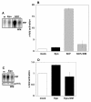
Strong activation of class Ia PI3 kinases by SCF, but PI3Kγ activation by Epo. Starved PEPs were mock-stimulated (m), stimulated with 0.3 U/ml Epo or with 25 ng/ml SCF with or without pretreatment with 100 nM WM as indicated. (A, B) The tyrosine-phosphorylated p85 regulatory subunits of activated class Ia PI3K enzymes (PI3Kα, β, δ) were immunoprecipitated from 500 μg total cell proteins with anti-phosphotyrosine mAb (4G10). To detect PI3K inositol kinase activity, immunoprecipitates were incubated with phosphatidylinositol (PI) and 32P-γ-ATP. Phosphatidylinositol phosphate (PIP) generated by active PI3Ks was separated from ATP by thin layer chromatography (TLC) and analyzed by phosphoimaging. A representative example of the results from the phosphoimaged experiments is shown in (A) and the quantitative analysis of the results obtained with PEPs from three different cord blood samples in (B). Epo and SCF significantly activate class I PI3Ks (pEpo < 0.01; pSCF < 0.001) and WM significantly inhibits the SCF-induced activation (p < 0.01). (C, D) Starved PEPs were mock stimulated (m), stimulated with 0.3 U/ml Epo with or without pretreatment with 100 nM WM as indicated. PI3 kinase activity was detected as in (A) and (B) but using anti-p110γ to immunoprecipitate the catalytic subunit of PI3Kγ. A representative example of the results from the phosphoimaged experiments is shown in (C) (upper panel) and the quantitative analysis of the results obtained with PEPs from three different cord blood samples in (D). Significant PI3Kγ activation (p < 0.001) and inhibition by WM (p < 0.001) was determined. Equal PI3Kγ immunoprecipitation was confirmed by western blot (C, lower panel).
Figure 4
Figure 4
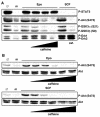
The PI3 kinase inhibitor caffeine inhibits Epo-induced Akt, GSK3α/β and Erk1/2 activation. Starved PEPs were mock-stimulated (m) or treated with 0.3 U/ml Epo or 25 ng/ml SCF as indicated. Some of the samples were pretreated with caffeine as detailed below. 100 μg cell protein were immunoblotted with P-STAT5, P-Akt, P-GSK3 α/β, P-Erk1/2 or Akt antibodies. Ø-lanes represent non-starved and non-treated PEPs. (A) Cells were pretreated with 0.1, 1 or 10 mM caffeine for 1 h before Epo stimulation and with 10 mM before SCF stimulation where indicated or (B) with 0.1, 0.3, 1, 3 or 10 mM caffeine for 1 h.
Figure 5
Figure 5
Activation of B-Raf by Epo is blocked by wortmannin. PEPs were mock-stimulated (m) or stimulated with 0.3 U/ml Epo or pretreated with 100 nM WM where indicated and then stimulated with Epo. (A, B) c-Raf1 was immunoprecipitated from 500 μg total cell protein with anti-c-Raf1. Precipitates were immunoblotted with anti-c-Raf1 for IP-control (A, lower panel) or incubated with GST-MEK and subsequently GST-ErkK63M and 32P-γ-ATP for coupled kinase assay. Proteins were separated by SDS-PAGE and phosphorylated GST-Erk1K63M analyzed by phosphoimaging. A representative example is shown in (A) (upper panel). Quantification of c-Raf1 activation from experiments with three different cord bloods is shown in B (pactivation < 0.001; pinhibition < 0.05). (C, D) B-Raf activation was analyzed as described in (A) and (B) but anti-B-Raf was used for immunoprecipitation and immunoblotting. Both activation and inhibition were statistically highly significant (pactivation < 0.001; pinhibition < 0.01).
Figure 6
Figure 6
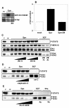
Inhibition of Erks by PKC inhibitors but not by Raf inhibitor. (A, B) Starved PEPs were mock-stimulated (m) or stimulated with 0.3 U/ml Epo with or without pretreatment with 10 μM ZM336372 (ZM) for 1 h. B-Raf was immunoprecipitated from 500 μg total cell protein with anti-B-Raf. Precipitates were immunoblotted with anti-B-Raf for IP-control (A, lower panel) or incubated with GST-MEK and subsequently GST-ErkK63M and 32P-γ-ATP for coupled kinase assay. Proteins were separated by SDS-PAGE and phosphorylated GST-Erk1K63M analyzed by phosphoimaging. A representative example is shown in (A) (upper panel). Quantification of B-Raf activation from experiments with three different cord bloods is shown in (B) (pactivation < 0.01; pinhibition < 0.05). (C) Starved PEPs were mock-stimulated (m) or pretreated with 0, 0.1, 1 or 10 μM ZM for 1 h or 4 h and stimulated with 0.3 U/ml Epo as indicated. Other cells were pretreated with 10 μM ZM and then stimulated with 25 ng/ml SCF. 100 μg total cell protein were immunoblotted with anti-P-STAT5, anti-P-MEK1/2, anti-Erk1/2 or anti-P-Erk1/2. Ø-lanes represent untreated PEPs. (D, E) Starved PEPs were mock-stimulated (m) or pretreated with PKC inhibitors and stimulated with 0.3 U/ml Epo or 25 ng/ml SCF. 100 μg of total cell protein were immunoblotted with anti-P-STAT5 and anti-P-Erk1/2. Inhibitor pretreatment was with 10, 100 or 1000 nM calphostin C for 1 h before Epo stimulation and with 1000 nM calphostin C before SCF stimulation. Other samples were pretreated with 0.1, 1, 10 or 100 μM Ro-31-8220 and stimulated with 0.3 U/ml Epo, or they were pretreated with 100 μM Ro-31-8220 where indicated and stimulated with 25 ng/ml SCF or 100 μM TPA for 10 min. 100 μg total cell protein were immunoblotted with anti-P-STAT5 or anti-P-Erk1/2. Ø-lanes represent untreated PEPs.
Figure 7
Figure 7
Schematic model of signaling events to MEKs and Erks induced by threshold concentrations of Epo. This basic Epo signal can be amplified or modulated by various other signaling pathways (not shown here) which become activated upon higher Epo concentrations and/or other factors and will often depend on SH2 domain interactions with the phosphorylated tyrosines in the cytoplasmic EpoR tail. PKCs could function as signal transducers for PI3Kγ, but it is also possible that PKCs are activated in a parallel pathway to PI3Kγ and that these two pathways converge to activate MEKs. B-Raf kinase does not significantly promote MAPK activation at low Epo concentrations, but since it is readily activated, it could play a role in signaling events induced by higher Epo concentrations.

Similar articles

Cited by

References

    1. Klingmuller U. The role of tyrosine phosphorylation in proliferation and maturation of erythroid progenitor cells--signals emanating from the erythropoietin receptor. Eur J Biochem. 1997;249:637–647. - PubMed
    1. Constantinescu SN, Ghaffari S, Lodish HF. The Erythropoietin Receptor: Structure, Activation and Intracellular Signal Transduction. Trends Endocrinol Metab. 1999;10:18–23. doi: 10.1016/S1043-2760(98)00101-5. - DOI - PubMed
    1. Wojchowski DM, Gregory RC, Miller CP, Pandit AK, Pircher TJ. Signal transduction in the erythropoietin receptor system. Exp Cell Res. 1999;253:143–156. doi: 10.1006/excr.1999.4673. - DOI - PubMed
    1. Lacombe C, Mayeux P. The molecular biology of erythropoietin. Nephrol Dial Transplant. 1999;14 Suppl 2:22–28. doi: 10.1093/ndt/14.suppl_2.22. - DOI - PubMed
    1. Digicaylioglu M, Lipton SA. Erythropoietin-mediated neuroprotection involves cross-talk between Jak2 and NF-kappaB signalling cascades. Nature. 2001;412:641–647. doi: 10.1038/35088074. - DOI - PubMed

MeSH terms