The spatial organization of the VirR boxes is critical for VirR-mediated expression of the perfringolysin O gene, pfoA, from Clostridium perfringens
- PMID: 15150217
- PMCID: PMC415773
- DOI: 10.1128/JB.186.11.3321-3330.2004
The spatial organization of the VirR boxes is critical for VirR-mediated expression of the perfringolysin O gene, pfoA, from Clostridium perfringens
Abstract
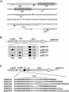
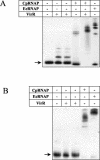
The transcriptional regulation of toxin production in the gram-positive anaerobe Clostridium perfringens involves a two-component signal transduction system that comprises the VirS sensor histidine kinase and its cognate response regulator, VirR. Previous studies showed that VirR binds independently to a pair of imperfect direct repeats, now designated VirR box 1 and VirR box 2, located immediately upstream of the promoter of the pfoA gene, which encodes the cholesterol-dependent cytolysin, perfringolysin O. For this study, we introduced mutated VirR boxes into a C. perfringens pfoA mutant and found that both VirR boxes are essential for transcriptional activation. Furthermore, the spacing between the VirR boxes and the distance between the VirR boxes and the -35 region are shown to be critical for perfringolysin O production. Other VirR boxes that were previously identified from the strain 13 genome sequence were also analyzed, with perfringolysin O production used as a reporter system. The results showed that placement of the different VirR boxes at the same position upstream of the pfoA promoter yields different levels of perfringolysin O activity. In all of these constructs, VirR was still capable of binding to the target DNA, indicating that DNA binding alone is not sufficient for transcriptional activation. Finally, we show that the C. perfringens RNA polymerase binds more efficiently to the pfoA promoter in the presence of VirR, indicating that interactions must occur between these proteins. We propose that these interactions are required for VirR-mediated transcriptional activation.
Figures


Similar articles
-
Identifying the Basis for VirS/VirR Two-Component Regulatory System Control of Clostridium perfringens Beta-Toxin Production.J Bacteriol. 2021 Aug 20;203(18):e0027921. doi: 10.1128/JB.00279-21. Epub 2021 Aug 20. J Bacteriol. 2021. PMID: 34228498 Free PMC article.
-
The VirR response regulator from Clostridium perfringens binds independently to two imperfect direct repeats located upstream of the pfoA promoter.J Bacteriol. 2000 Jan;182(1):57-66. doi: 10.1128/JB.182.1.57-66.2000. J Bacteriol. 2000. PMID: 10613863 Free PMC article.
-
The virR/virS locus regulates the transcription of genes encoding extracellular toxin production in Clostridium perfringens.J Bacteriol. 1996 May;178(9):2514-20. doi: 10.1128/jb.178.9.2514-2520.1996. J Bacteriol. 1996. PMID: 8626316 Free PMC article.
-
Gene regulation by the VirS/VirR system in Clostridium perfringens.Anaerobe. 2016 Oct;41:5-9. doi: 10.1016/j.anaerobe.2016.06.003. Epub 2016 Jun 11. Anaerobe. 2016. PMID: 27296833 Review.
-
Regulation of toxin gene expression in Clostridium perfringens.Res Microbiol. 2015 May;166(4):280-9. doi: 10.1016/j.resmic.2014.09.010. Epub 2014 Oct 7. Res Microbiol. 2015. PMID: 25303832 Review.
Cited by
-
Host cell-induced signaling causes Clostridium perfringens to upregulate production of toxins important for intestinal infections.Gut Microbes. 2014 Jan-Feb;5(1):96-107. doi: 10.4161/gmic.26419. Epub 2013 Sep 10. Gut Microbes. 2014. PMID: 24061146 Free PMC article. Review.
-
Identifying the Basis for VirS/VirR Two-Component Regulatory System Control of Clostridium perfringens Beta-Toxin Production.J Bacteriol. 2021 Aug 20;203(18):e0027921. doi: 10.1128/JB.00279-21. Epub 2021 Aug 20. J Bacteriol. 2021. PMID: 34228498 Free PMC article.
-
plcR papR-independent expression of anthrolysin O by Bacillus anthracis.J Bacteriol. 2006 Nov;188(22):7823-9. doi: 10.1128/JB.00525-06. Epub 2006 Sep 15. J Bacteriol. 2006. PMID: 16980467 Free PMC article.
-
Clostridium Perfringens Toxins Involved in Mammalian Veterinary Diseases.Open Toxinology J. 2010;2:24-42. Open Toxinology J. 2010. PMID: 24511335 Free PMC article.
-
The VirSR two-component signal transduction system regulates NetB toxin production in Clostridium perfringens.Infect Immun. 2010 Jul;78(7):3064-72. doi: 10.1128/IAI.00123-10. Epub 2010 May 10. Infect Immun. 2010. PMID: 20457789 Free PMC article.
References
-
- Awad, M. M., A. E. Bryant, D. L. Stevens, and J. I. Rood. 1995. Virulence studies on chromosomal α-toxin and θ-toxin mutants constructed by allelic exchange provide genetic evidence for the essential role of α-toxin in Clostridium perfringens-mediated gas gangrene. Mol. Microbiol. 15:191-202. - PubMed
-
- Awad, M. M., and J. I. Rood. 1997. Isolation of α-toxin, θ-toxin and κ-toxin mutants of Clostridium perfringens by Tn916 mutagenesis. Microb. Pathog. 22:275-284. - PubMed
-
- Bannam, T. L., and J. I. Rood. 1993. Clostridium perfringens-Escherichia coli shuttle vectors that carry single antibiotic resistance determinants. Plasmid 29:223-235. - PubMed
Publication types
MeSH terms
Substances
LinkOut - more resources
Full Text Sources

