. 2004 Jun 1;101(22):8479-84.
doi: 10.1073/pnas.0401000101. Epub 2004 May 18.

Completion of neuronal migration regulated by loss of Ca(2+) transients

Affiliations

Completion of neuronal migration regulated by loss of Ca(2+) transients

Tatsuro Kumada et al. Proc Natl Acad Sci U S A. .

Abstract

The migration of immature neurons constitutes one of the major processes by which the central nervous system takes shape. Completing the migration at the final destination requires the loss of cell body motility, but little is known about the signaling mechanisms underlying this process. Here, we show that a loss of transient Ca(2+) elevations triggers the completion of cerebellar granule cell migration. Simultaneous observation of the intracellular Ca(2+) levels and cell movement in cerebellar slices of the early postnatal mice revealed that granule cells exhibit distinct frequencies of the transient Ca(2+) elevations as they migrate in different cortical layers, and complete the migration only after the loss of Ca(2+) elevations. The reduction of the Ca(2+) elevation frequency by decreasing Ca(2+) influx, or by inhibiting the activity of phospholipase C, PKC, or Ca(2+)/calmodulin, halted the granule cell movement prematurely. In contrast, increasing the Ca(2+) elevation frequency by increasing Ca(2+) release from internal stores, or by elevating intracellular cAMP levels, significantly delayed the completion of granule cell migration. The timing of the loss of Ca(2+) elevations was intrinsically set in the granule cells and influenced by external cues. These results suggest that Ca(2+) signaling, dictated by multiple signaling systems, functions as a mediator for completing the migration of immature neurons.


Figures
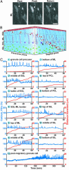
Fig. 1.
Fig. 1.
Dynamic changes in Ca2+ transient frequency of granule cells along the migratory pathway. (A) Typical example of a migrating granule cell in the molecular layer showing transient elevations of the intracellular Ca2+ levels in its soma (indicated by white arrows). (Bar = 7 μm.) (B) Schematic diagram showing the sequence of cerebellar granule cell migration from the birthplace at the top of the EGL to the final destination at the bottom of the IGL. The numbers 1–13 represent the position of granule cells along the migratory pathway and the stage of the differentiation in order: 1, granule cell precursor at the top of the EGL; 2–4, tangential migration at the middle and bottom of the EGL; 5–7, radial migration along the Bergmann glial process in the molecular layer; 8 and 9, stationary period in the Purkinje cell layer; 10 and 11, radial migration at the top and middle of the IGL; 12, completion of migration at the bottom of the IGL; and 13, postmigratory granule cell at the bottom of the IGL. B, Bergmann glial cell; P, Purkinje cell. (C) Sequential changes in the Ca2+ transients (blue lines) and the distance traversed by granule cells (red lines) over time. The number at the top of each graph corresponds to the number in B. Upward deflections in blue lines represent elevations of Ca2+ levels, and downward deflections indicate decreases of Ca2+ levels.
Fig. 2.
Fig. 2.
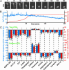
Relationship between the Ca2+ transient frequency and the granule cell position along the migratory pathway (Upper) and between the migration rate and the granule cell position (Lower). Each column represents the average values and SD obtained from >30 granule cells.
Fig. 3.
Fig. 3.
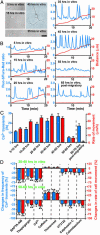
Completion of granule cell migration triggered by loss of Ca2+ transients. (A) Time-lapse images showing a typical example of the completion of granule cell migration at the bottom of the IGL. Elapsed time (in minutes) is indicated on the top of each photograph. (Bar = 10 μm.) (B) Alterations of spontaneous Ca2+ transients in the granule cell soma before and after the completion of migration. Changes in Ca2+ levels (blue line) and distance (red line) traversed by the granule cell shown in A are plotted as a function of elapsed time. (C) Effects of the changes in the Ca2+ transient frequency on the migration rate of granule cells at the top and bottom of the IGL. We chose the granule cells that migrated at the upper part of the top and bottom area, where the majority of the cells moved toward the IGL–white matter border at the average rates of ≈11 μm/h (top) and ≈4 μm/h (bottom). BAPTA-AM (10 μM), CdCl2 (500 μM), d-2-amino-5-phosphonopentanoic acid (100 μM), thapsigargin (1 μM), caffeine (10 mM), thimerosal (5 μM), U73122 (1 μM), calphostin C (100 nM), calmidazolium (5 μM), forskolin (30 μM), or somatostatin-14 (SST-14, 1 μM) was added to the medium in separate experiments, or extracellular Ca2+ concentrations were lowered from 1.8 mM to 0.1 mM after 60-min control observations. Here and in Fig. 4D, the effects of each treatment were evaluated by dividing the number of Ca2+ transients and the distance traveled during the 60 min after the application of each reagent by the number of Ca2+ transients and the distance traveled during the first 60 min in the absence of each reagent. Each column represents the average changes (obtained from >30 cells) in the number of Ca2+ transients (blue) or the migration rate (red). *, P < 0.05; **, P < 0.01.
Fig. 4.
Fig. 4.
Loss of the Ca2+ transients in isolated granule cells set by intrinsic programs. (A) Photomicrographs of isolated granule cells in the microexplant culture of the early postnatal cerebellum. (Bar = 12 μm.) (B) Changes in the Ca2+ transients and the movement of isolated granule cells as elapse time in vitro goes by. The Ca2+ levels (blue lines) and the distance (red lines) traversed are plotted as a function of elapsed time. Upward deflections in blue lines represent the elevations of the intracellular Ca2+ levels, and downward deflections indicate the decreases of the intracellular Ca2+ levels. (C) Sequential changes in the Ca2+ transient frequency and the migration rate of isolated granule cells in the microexplant culture. Each column represents the average values (obtained from >30 granule cells) of Ca2+ transients (blue) and the migration rate (red). (D) Effects of the changes in the Ca2+ transient frequency on the migration rate of isolated granule cells at 30–40 h in vitro and 50–60 h in vitro. BAPTA-AM (10 μM), thapsigargin (1 μM), CdCl2 (500 μM), caffeine (10 mM), thimerosal (5 μM), U73122 (1 μM), calphostin C (100 nM), and calmidazolium (5 μM) were added to the culture medium in separate experiments after 60-min control observations. Each column represents the average changes (obtained from >30 cells) in the Ca2+ transient frequency (blue) or the migration rate (red). *, P < 0.05; **, P < 0.01.

Similar articles

Cited by

References

    1. Rakic, P. (1990) Experientia 46, 882-891. - PubMed
    1. Hatten, M. E. (1999) Annu. Rev. Neurosci. 22, 511-539. - PubMed
    1. Caviness, V. S., Jr., & Rakic, P. (1978) Annu. Rev. Neurosci. 1, 297-326. - PubMed
    1. Ross, M. E. & Walsh, C. A. (2001) Annu. Rev. Neurosci. 24, 1041-1070. - PubMed
    1. Rakic, P. (1971) J. Comp. Neurol. 141, 283-312. - PubMed

Publication types