. 2004 Jun 1;101(22):8455-60.
doi: 10.1073/pnas.0400576101. Epub 2004 May 18.

Severe acute respiratory syndrome coronavirus (SARS-CoV) infection inhibition using spike protein heptad repeat-derived peptides

Affiliations

Severe acute respiratory syndrome coronavirus (SARS-CoV) infection inhibition using spike protein heptad repeat-derived peptides

Berend Jan Bosch et al. Proc Natl Acad Sci U S A. .

Abstract

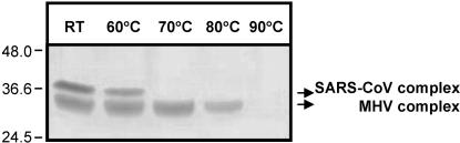
The coronavirus SARS-CoV is the primary cause of the life-threatening severe acute respiratory syndrome (SARS). With the aim of developing therapeutic agents, we have tested peptides derived from the membrane-proximal (HR2) and membrane-distal (HR1) heptad repeat region of the spike protein as inhibitors of SARS-CoV infection of Vero cells. It appeared that HR2 peptides, but not HR1 peptides, were inhibitory. Their efficacy was, however, significantly lower than that of corresponding HR2 peptides of the murine coronavirus mouse hepatitis virus (MHV) in inhibiting MHV infection. Biochemical and electron microscopical analyses showed that, when mixed, SARS-CoV HR1 and HR2 peptides assemble into a six-helix bundle consisting of HR1 as a central triple-stranded coiled coil in association with three HR2 alpha-helices oriented in an antiparallel manner. The stability of this complex, as measured by its resistance to heat dissociation, appeared to be much lower than that of the corresponding MHV complex, which may explain the different inhibitory potencies of the HR2 peptides. Analogous to other class I viral fusion proteins, the six-helix complex supposedly represents a postfusion conformation that is formed after insertion of the fusion peptide, proposed here for coronaviruses to be located immediately upstream of HR1, into the target membrane. The resulting close apposition of fusion peptide and spike transmembrane domain facilitates membrane fusion. The inhibitory potency of the SARS-CoV HR2-peptides provides an attractive basis for the development of a therapeutic drug for SARS.


Figures

Fig. 1.
Fig. 1.
(A) Schematic representation of the coronavirus spike protein structure. The glycoprotein has an N-terminal signal sequence (SS) and a transmembrane domain (TM) close to the C terminus. Group 2 and 3 coronavirus spike proteins are proteolytically cleaved (arrow) in an S1 and an S2 subunit, which are noncovalently linked. S2 contains two heptad repeat regions (shaded bars), HR1 and HR2, as indicated. (B) clustalw multiple sequence alignment of coronavirus spike proteins. Shown is the alignment of the HR1 and HR2 domains of the recently identified SARS-CoV (strain TOR2) with those of the group 1 coronaviruses FIPV (feline infectious peritonitis virus strain 79–1146) and HCoV-229E (human coronavirus strain 229E), the group 2 coronaviruses MHV-A59 (mouse hepatitis virus strain A59) and HCoV-OC43 (human coronavirus strain OC43), and the group 3 coronavirus IBV (infectious bronchitis virus strain Beaudette) (GenBank accession nos. P59594, VGIH79, VGIHHC, P11224, CAA83661, and P11223, respectively). Dark shading marks sequence identity, whereas lighter shading represents sequence similarity. The alignment shows a remarkable insertion of exactly two heptad repeats (14 aa) in both HR1 and HR2 of HCV-229E and FIPV, a characteristic of all group 1 viruses. The predicted hydrophobic heptad repeat “a” and “d” residues are indicated above the sequence. Asterisks denote conserved residues, dots represent similar residues. The amino acid sequences of the HR1-derived peptides HR1, HR1a, HR1b, HR1c, and a FLAG-tagged HR1 (Fl.HR1) and of the HR2 derived peptides HR2, HR2-1 and a FLAG-tagged HR2 (Fl.HR2) of SARS-CoV used in this study are presented in italics below the alignments. N-terminal glycine and serine residues derived from the thrombin proteolytic cleavage site of the GST fusion protein are in parentheses.
Fig. 2.
Fig. 2.
Inhibition of SARS-CoV infection by HR peptides. (A) VERO cells were mock infected or infected with SARS-CoV (multiplicity of infection = 0.5) in the presence of the HR2-1 peptide (sHR2-1) at concentrations of 0, 5, or 25 μM and incubated in medium containing the same concentration of peptide. An infection in the presence of peptide (25 μM) corresponding to the HR2 domain of MHV (mHR2) was taken along as a negative control. At 16 h after infection, cells were fixed, and SARS-CoV-positive cells were visualized by immunofluorescence staining.
Fig. 3.
Fig. 3.
Complex formation of SARS-CoV HR1 and HR2 peptides. Comparison of SARS-CoV and MHV. HR1 and HR2 peptides on their own or as a preincubated equimolar mixture were subjected to 15% Tricine SDS/PAGE. Just before loading onto the gel, some samples were heated at 100°C. In Left and Right, complex formation was analyzed after a 3 h and an O/N incubation, respectively.
Fig. 4.
Fig. 4.
Stoichiometry of peptides in HR1–HR2 complexes. (A) Size-exclusion HPLC elution profile of the HR1 peptide (dotted line), HR2 peptide (dashed line), and an O/N preincubated equimolar mix of the HR1 and HR2 peptides (solid line). (B) Nano-ESI-TOF mass spectrum of the HR1 and HR2 peptides and of the HR1–HR2 complex. m/z values of the peaks of the HR1 (+; Top), the HR2 peptide (-; Middle) or the HR1–HR2 peptide complex (±±±; Bottom) are indicated, with the charge state assignment in brackets. The convoluted average molecular mass (Mw) of the HR1 and HR2 peptides and of the HR1–HR2 peptide complex are indicated as well. Peaks corresponding to the HR1 and HR2 monomers are visible in the lower m/z range of the spectrum of the complex, indicating that a fraction of it dissociated during the procedure.
Fig. 5.
Fig. 5.
Comparative temperature stabilities of HR1–HR2 complexes of SARS-CoV and MHV. Equal amounts of SARS-CoV and MHV HR1–HR2 complexes were pooled, subsequently incubated for 5 min at the indicated temperatures in 1× Tricine sample buffer and analyzed directly by SDS/PAGE in a 15% Tricine gel. Positions of the HR1–HR2 complex of SARS-CoV and MHV are indicated on the right, and the molecular mass markers are indicated at the left.

Similar articles

Cited by

References

    1. Gallagher, T. M. & Buchmeier, M. J. (2001) Virology 279, 371-374. - PMC - PubMed
    1. Bosch, B. J., van der Zee, R., de Haan, C. A. & Rottier, P. J. (2003) J. Virol. 77, 8801-8811. - PMC - PubMed
    1. Lescar, J., Roussel, A., Wien, M. W., Navaza, J., Fuller, S. D., Wengler, G. & Rey, F. A. (2001) Cell 105, 137-148. - PubMed
    1. Eckert, D. M. & Kim, P. S. (2001) Annu. Rev. Biochem. 70, 777-810. - PubMed
    1. Delmas, B. & Laude, H. (1990) J. Virol. 64, 5367-5375. - PMC - PubMed

MeSH terms

LinkOut - more resources