. 2004 Jun 14;90(12):2390-6.
doi: 10.1038/sj.bjc.6601838.

Alteration of the copy number and deletion of mitochondrial DNA in human hepatocellular carcinoma

Affiliations

Alteration of the copy number and deletion of mitochondrial DNA in human hepatocellular carcinoma

P H Yin et al. Br J Cancer. .

Abstract

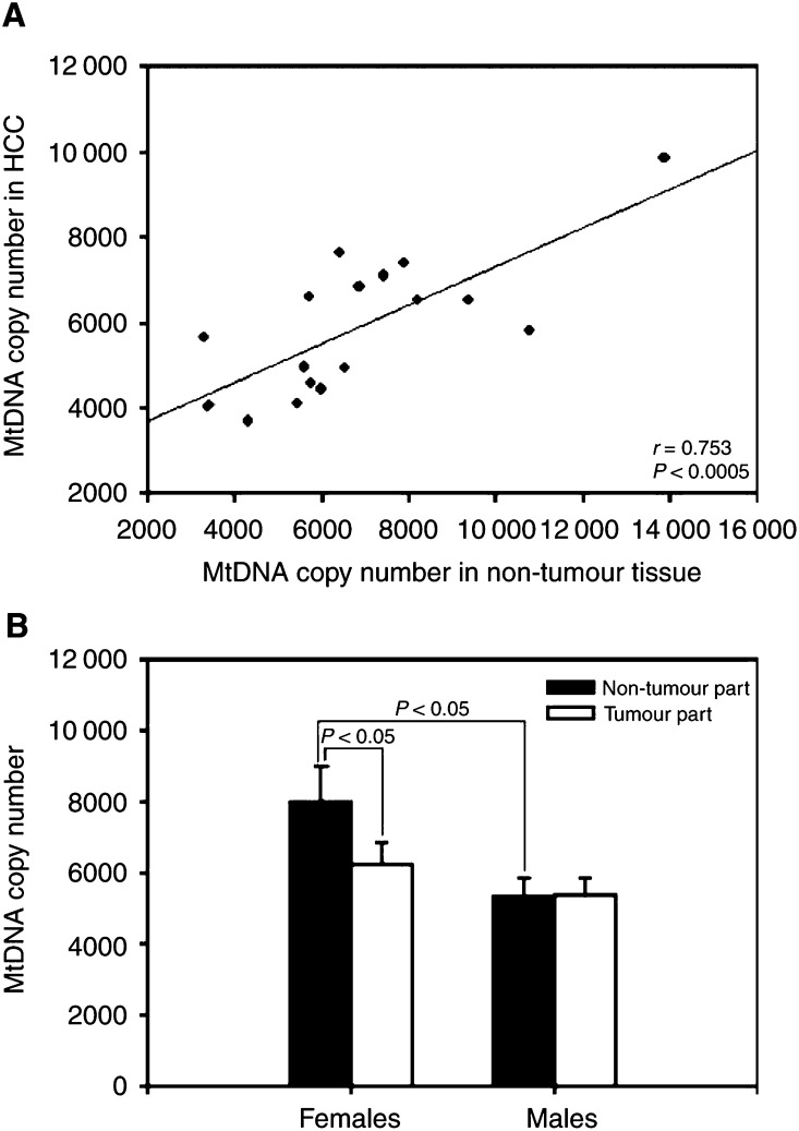
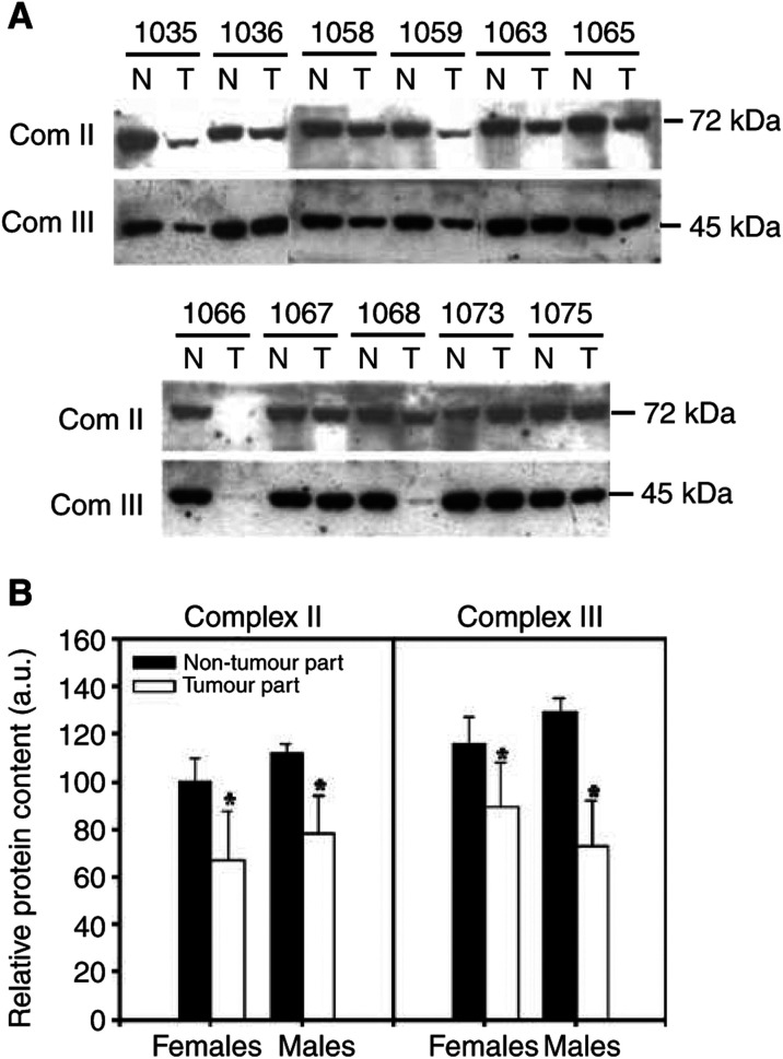
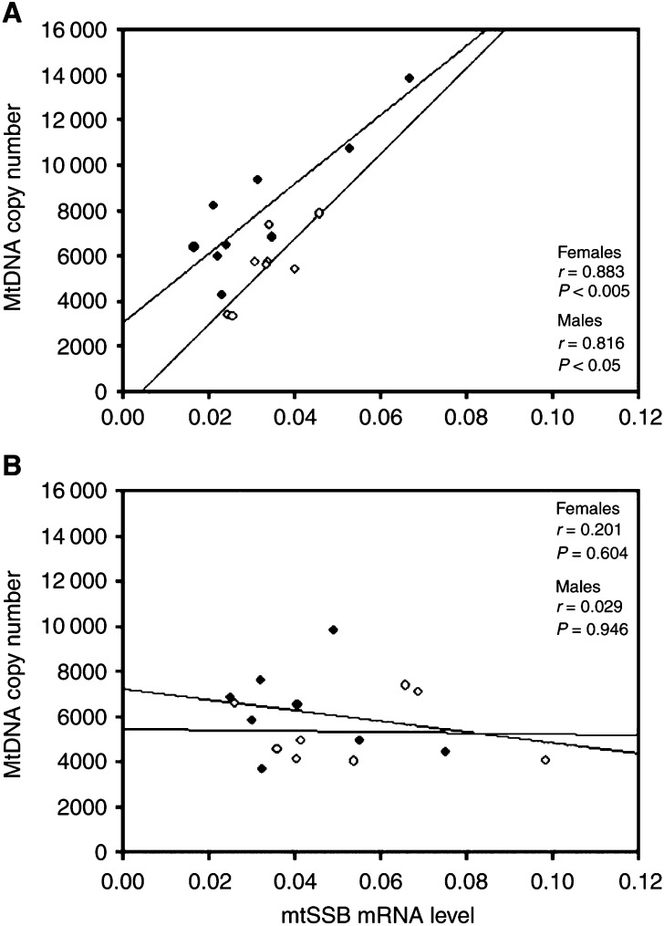
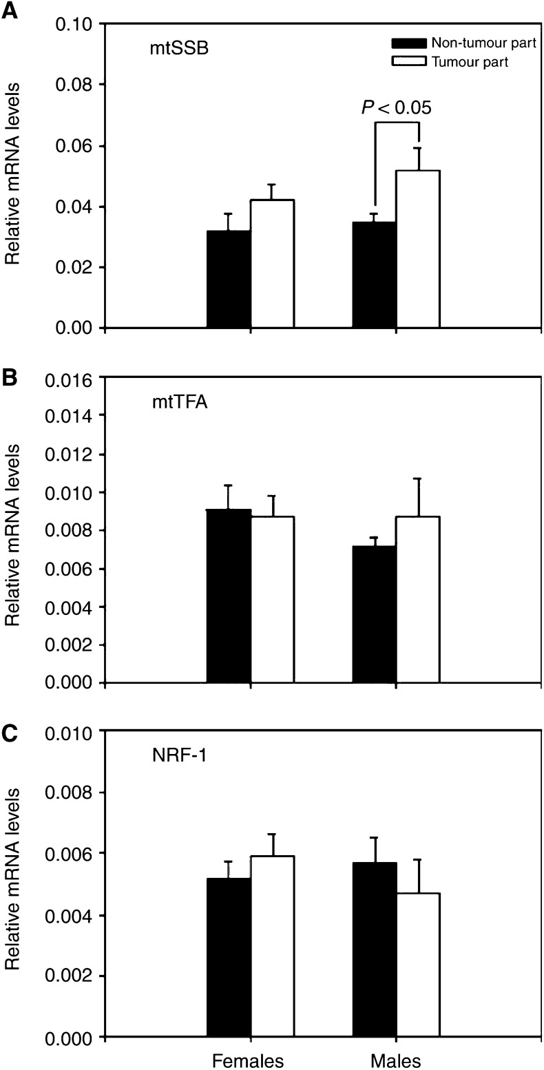
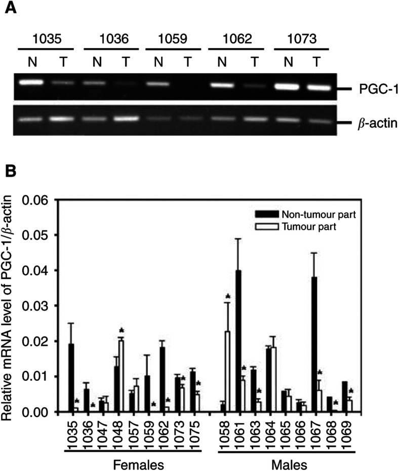
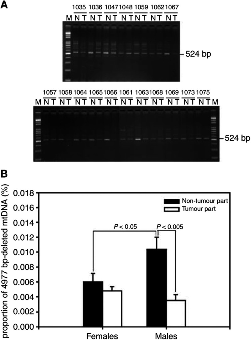
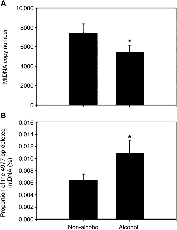
Somatic mutations in mitochondrial DNA (mtDNA) have been detected in hepatocellular carcinoma (HCC). However, it remains unclear whether mtDNA copy number and mitochondrial biogenesis are altered in HCC. In this study, we found that mtDNA copy number and the content of mitochondrial respiratory proteins were reduced in HCCs as compared with the corresponding non-tumorous livers. MtDNA copy number was significantly reduced in female HCC but not in male HCC. Expression of the peroxisome proliferator-activated receptor gamma coactivator-1 was significantly repressed in HCCs (P<0.005), while the expression of the mitochondrial single-strand DNA-binding protein was upregulated, indicating that the regulation of mitochondria biogenesis is disturbed in HCC. Moreover, 22% of HCCs carried a somatic mutation in the mtDNA D-loop region. The non-tumorous liver of the HCC patients with a long-term alcohol-drinking history contained reduced mtDNA copy number (P<0.05) and higher level of the 4977 bp-deleted mtDNA (P<0.05) as compared with non-alcohol patients. Our results suggest that reduced mtDNA copy number, impaired mitochondrial biogenesis and somatic mutations in mtDNA are important events during carcinogenesis of HCC, and the differential alterations in mtDNA of male and female HCC may contribute to the differences in the clinical manifestation between female and male HCC patients.


Figures

Figure 1
Figure 1
MtDNA copy number in HCC. Panel A shows a positive correlation between the copy number of mtDNA within the tumour tissues and the corresponding non-tumorous tissues (r=0.753; P<0.0005). Panel B shows the mean copy number of mtDNA within the tumour tissues and the corresponding non-tumorous tissues from the female and male HCC patients. The results are presented as mean±s.e.m. Comparisons between the group means were analysed by Student's t-test.
Figure 2
Figure 2
Mitochondrial respiratory proteins in HCC. Panel A shows the relative content of the mitochondrial respiratory proteins, the 72 kDa subunit of the succinate-ubiquinol oxidoreductase (complex II) and the core 2 subunit of the ubiquinol-cytochrome c oxidoreductase (complex III), determined by immunoblot analysis described in Materials and methods. Panel B shows the histogram of the protein content (expressed as arbitrary units, a.u.) of each mitochondrial respiratory protein in non-tumorous and tumour tissues from five female and six male HCC patients. The results are presented as mean±s.e.m. * represents a significant decrease in the protein content of the tumour tissues as compared with that of the corresponding non-tumorous tissues (P<0.05, Student's t-test).
Figure 3
Figure 3
Correlation between the expression of mtSSB mRNA and mtDNA copy number. Panel A shows a positive correlation between the expression of mtSSB mRNA and the copy number of mtDNA within the non-tumorous tissues of the female HCC patients (the closed symbols, r=0.883; P<0.005) and of the male HCC patients (the open symbols, r=0.816; P<0.05), respectively. Panel B shows a loss of the relationship between the expression of mtSSB mRNA and the copy number of mtDNA within the tumour tissues of the female HCC patients (the closed symbols, r=0.201; P=0.604) and of the male HCC patients (the open symbols, r=0.029; P=0.946), respectively.
Figure 4
Figure 4
Alterations in the mRNA expressions of mtSSB, mtTFA and NRF-1 in HCC. Alterations in the mRNA expressions of mtSSB (panel A), mtTFA (panel B) and NRF-1 (panel C) were analysed by the ΔΔCt method, as described in Materials and methods. Data are first normalised to the level of β-actin in each sample. The results are presented as mean±s.e.m. Comparisons between the group means were analysed by Student's t-test. A value of P<0.05 was considered statistically significant.
Figure 5
Figure 5
Alteration in the mRNA expression of PGC-1 in HCC. Alteration in the mRNA expression of PGC-1 was analysed by RT–PCR method (panel A) and was quantified by the ΔΔCt method (panel B) as described in Materials and methods. Data are first normalised to the level of β-actin in each sample. The results are presented as mean±s.e.m. * represents a significant alteration in the mRNA level of PGC-1 of the tumour tissues as compared with that of the corresponding non-tumorous tissues (P<0.05, Student's t-test).
Figure 6
Figure 6
MtDNA 4977 bp deletion in HCC MtDNA with the 4977 bp deletion was detected by the PCR technique described in Materials and methods. Using the primers L8150 and H13650, a 524 bp PCR product amplified from the 4977 bp-deleted mtDNA in the tumour part (T) and in the corresponding non-tumorous tissue (N) were separated on a 1.5% agarose gel and detected under UV transillumination after ethidium bromide staining. M: 100 bp DNA ladder. The numbers above the gel lanes indicate the patients (panel A). The mean proportions of the 4977 bp-deleted mtDNA in the tumour tissues were compared with the corresponding non-tumour tissues (panel B). The results are presented as mean±s.e.m. Comparisons between the group means were analysed by Student's t-test.
Figure 7
Figure 7
The effect of alcohol consumption on the level of mtDNA copy number and the accumulation of the 4977 bp-deleted mtDNA in the liver tissues. The level of mtDNA copy number (A) and the proportion of the 4977 bp-deleted mtDNA (B) were, respectively, determined by the PCR techniques described in Materials and methods. The mean copy number of mtDNA in the non-tumorous tissues of seven HCC patients with a history of long-term drinking of alcohol (alcohol group) was significantly lower than that of 11 HCC patients without a history of long-term drinking of alcohol (non-alcohol group) (*P<0.05, Student's t-test). In contrast, the mean proportion of the 4977 bp-deleted mtDNA in the non-tumorous tissues of the alcohol group was significantly higher than that of the non-alcohol group (*P<0.05, Student's t-test).

Similar articles

Cited by

References

    1. Anderson S, Bankier AT, Barrell BG, De Bruijn MHL, Coulson AR, Drouin J, Eperon IC, Nierlich DP, Roe BA, Sanger F, Schreier PH, Smith AJH, Staden R, Young IG (1981) Sequence and organization of the human mitochondrial genome. Nature 290: 457–465 - PubMed
    1. Augenlicht LH, Heerdt BG (2001) Mitochondria: integrators in tumorigenesis? Nat Genet 28: 104–105 - PubMed
    1. Cederbaum AI (1999) Effects of alcohol on hepatic mitochondrial function and DNA. Gastroenterology 117: 265–269 - PubMed
    1. Chen CJ, Yu MW, Liaw YF (1997) Epidemiological characteristics and risk factors of hepatocellular carcinoma. J Gastroenterol Hepatol 12: S294–S308 - PubMed
    1. Cuezva JM, Krajewska M, De Heredia ML, Krajewski S, Santamaria G, Kim H, Zapata JM (2002) The bioenergetic signature of cancer: a marker of tumor progression. Cancer Res 62: 6674–6681 - PubMed

Publication types