. 2004 May 19;24(20):4737-48.
doi: 10.1523/JNEUROSCI.0649-04.2004.

Cholecystokinin modulates migration of gonadotropin-releasing hormone-1 neurons

Affiliations

Cholecystokinin modulates migration of gonadotropin-releasing hormone-1 neurons

Paolo Giacobini et al. J Neurosci. .

Abstract

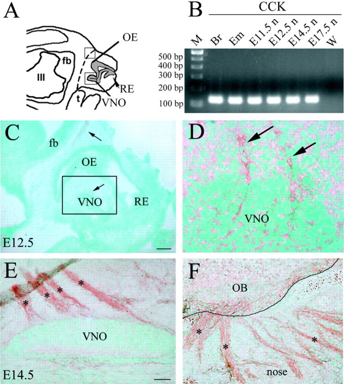
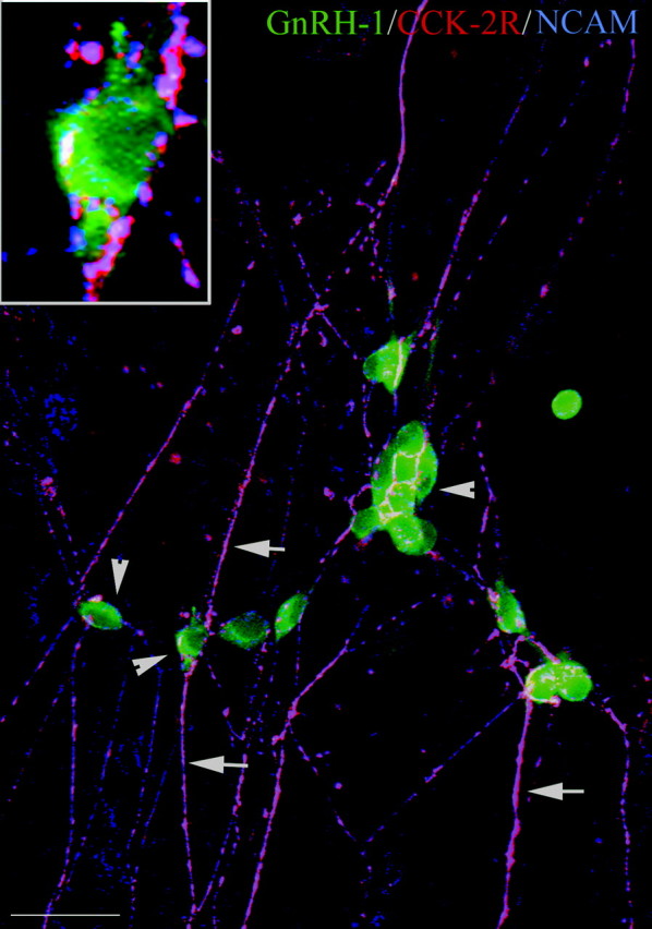
Expression of the brain-gut peptide cholecystokinin (CCK) in the developing olfactory-gonadotropin-releasing hormone-1 (GnRH-1) neuroendocrine systems was characterized, and the function of CCK in these systems was analyzed both in vivo and in vitro. We present novel data demonstrating that CCK transcript and protein are expressed in sensory cells in the developing olfactory epithelium and vomeronasal organ, with both ligand and receptors (CCK-1R and CCK-2R) found on olfactory axons throughout prenatal development. In addition, migrating GnRH-1 neurons in nasal regions express CCK-1R but not CCK-2R receptors. The role of CCK in olfactory-GnRH-1 system development was evaluated using nasal explants, after assessing that the in vivo expression of both CCK and CCK receptors was mimicked in this in vitro model. Exogenous application of CCK (10(-7) m) reduced both olfactory axon outgrowth and migration of GnRH-1 cells. This inhibition was mediated by CCK-1R receptors. Moreover, CCK-1R but not CCK-2R antagonism caused a shift in the location of GnRH-1 neurons, increasing the distance that the cells migrated. GnRH-1 neuronal migration in mice carrying a genetic deletion of either CCK-1R or CCK-2R receptor genes was also analyzed. At embryonic day 14.5, the total number of GnRH-1 cells was identical in wild-type and mutant mice; however, the number of GnRH-1 neurons within forebrain was significantly greater in CCK-1R-/- embryos, consistent with an accelerated migratory process. These results indicate that CCK provides an inhibitory influence on GnRH-1 neuronal migration, contributing to the appropriate entrance of these neuroendocrine cells into the brain, and thus represent the first report of a developmental role for CCK.


Figures

Figure 1.
Figure 1.
Olfactory system expresses CCK during embryonic development. A, Schematic of an E12.5 head; forebrain (fb), olfactory epithelium (OE), presumptive vomeronasal organ (VNO), respiratory epithelium (RE), tongue (t), and third ventricle (III) are depicted. Dashed line indicates boundary between nose and brain and represents region taken for nasal RNA isolation in B. B, Gel documentation of products produced by RT-PCR amplification using nested CCK primers. Total RNA was isolated from nose (E11.5n–E17.5n). Adult brain (Br) and E17.5 whole embryo (Em) were used as positive control tissues. A band of expected size (169 bp) was detected in positive control tissues after first-round PCR (data not shown). Second-round PCR, using 1 μl of first-round amplicons and second set of nested primers, yielded the predicted 135 bp product in all samples but water [w; a DNA ladder of 100 bp markers (m) was loaded in left lane]. C, Sagittal section of an E12.5 mouse embryo (16 μm) immunostained for CCK. CCK-immunopositive tracks extended from the presumptive VNO (D, see boxed area and high magnification), and a light signal was detected at the boundaries between the nasal region and the forebrain (nasal–forebrain junction; arrow). E, F, Immunohistochemistry for CCK on a sagittal section of an E14.5 embryo. At this stage, a notable increase in CCK immunoreactivity was evident throughout the axon bundles emerging from the VNO (E, asterisks) and the olfactory axons reaching the nasal–forebrain junction (F, asterisks). OB, Olfactory bulb. Scale bars: C, 150 μm; D, 15 μm; E, F, 50 μm.
Figure 7.
Figure 7.
CCK alters olfactory axon outgrowth and GnRH-1 cell migration. A, Photomicrograph of nasal explant immunocytochemically labeled for GnRH-1 (brown) and peripherin (blue), at 5 DIV. Many GnRH-1 neurons were visualized within the main tissue mass and in the periphery. Images were digitized and overlaid to a calibration meter composed of concentric arcs. Olfactory axon outgrowth (left histograms) as well as front of GnRH-1 cell migration (right histograms) were calculated as a function of treatment. GnRH-1 neurons remained apposed to peripherin fibers (along which they migrate) under all conditions. B, C, CCK (100 nm) applied at 3 DIV significantly decreased, at 5 DIV, olfactory axon outgrowth (B) and the front of migration of GnRH-1 cells (C) compared with controls (significantly different from control; ap < 0.001). CCK was ineffective in the presence of the CCK-1R-selective antagonist (lorglumide 100 nm; significantly different from CCK; bp < 0.05). Such treatment returned both parameters to control values. Olfactory axon outgrowth and the front of migration were similar in explants treated with CCK versus CCK in the presence of the CCK-2R antagonist (L-365, 260, 100 nm). D, E, Endogenous CCK is functionally active in nasal explants and acts via CCK-1R. CCK-1R antagonist treatment did not alter axonal outgrowth (D) but significantly increased the front of GnRH-1 cell migration (E) (significantly different from control and CCK-2R antagonist treated groups; abp< 0.01). CCK-2R selective antagonist application did not alter either olfactory axon outgrowth (D) or the front of migration (E) compared with controls. Scale bars: A, 200 μm; inset, 15 μm.
Figure 8.
Figure 8.
Migration of GnRH-1 neurons in CCK-1R–/– mutant embryos is accelerated. Quantitation of GnRH-1 cell number in three regions along the migratory pathway within sagittal and coronal sections from E14.5 WT, CCK-1R KO, and CCK-2R KO mice was performed. A, B, Photomicrographs showing GnRH-1 (brown)–peripherin (blue) immunoreactivity in a sagittal section of a CCK-1R KO embryo. The areas of analysis for GnRH-1 neuron location along the migratory pathway are shown in A. The boxed area = nasal–forebrain junction (N/FJ); cells located in areas outside this region were classified as in either the CNS (B) or nose. C, Analysis of GnRH-1 neuron location revealed an increase in the number of GnRH-1 neurons in the CNS of CCK-1R KO compared with WT and CCK-2R KO mice (**p < 0.01; by post hoc Fisher's LSD). Consistent with these data, fewer (although it did not reach significance; Fisher's LSD post hoc test; p=0.07) GnRH-1 cells were located in the N/FJ area of CCK-1R mutant embryos compared with WT and CCK-2R-deficient mice. Scale bars: A, 150 μm; B, 50 μm.
Figure 2.
Figure 2.
Olfactory sensory neurons express CCK mRNA and protein during development. A, Schematic of E17.5 coronal section depicting location of VNO and OE. NMC, Nasal midline cartilage. B, C, Immunocytochemistry for CCK on E17.5 coronal mouse sections. At E17.5, olfactory receptor neurons were CCK positive (B). Perikarya (arrows; outlined enlarged picture), apical dendrites (arrowheads), and axons of these neurons (data not shown) were stained for CCK. In the VNO (C), CCK was expressed in a small population of sensory neurons, toward the central region of VNO neuroepithelium. This CCK labeling was in cell soma (inset) and occasionally in apical dendrites. D, Photographs of E14.5 and E17.5 sagittal sections after LCM. The representative pictures show two examples of microdissected OE (leftpanel) and pituitary (Pit; right panel). Total RNA isolated from dissected region of OE and VNO was subjected to RT-PCR followed by two PCR rounds using nested primers. A fragment of the expected size (135 bp) was detected for CCK in the OE but not in VNO or water at E14.5 (left panel). Expression of the olfactory marker, EBF-2 (165 bp), and β-tubulin (data not shown) confirmed the morphology of the dissected tissue. Total RNA isolated from dissected E17.5 OE, VNO, and Pit was processed as described for E14.5 tissue. A strong CCK band was evident in OE and Pit, whereas a weak but detectable product was present in VNO. No PCR product was observed in reactions that omitted either reverse transcriptase (data not shown) or starting material (water). PCR analysis using EBF-2 primers showed expected amplicons in OE and VNO but not in Pit (right panel). E, Sagittal section of an E14.5 mouse nose stained for GnRH-1 and peripherin (intermediate filament protein expressed in olfactory–vomeronasal axons). GnRH-1 neurons (blue staining) migrate along peripherin-positive axons (brown staining) through the nasal mesenchyme. Inset shows a high magnification of GnRH-1 neurons migrating out of the VNO (arrows indicate GnRH-1 cells associated with peripherin-immunoreactive fibers). F, Immunocytochemistry of an E14.5 mouse using GnRH-1 and CCK antisera. GnRH-1 neurons (green) emerge from the VNO and migrate along CCK-positive axons (red). Note that staining pattern resembles that in GnRH-1–peripher in double labeling (E). Scale bars: B, 10 μm; inset in B, 5 μm; C, 25 μm; inset in C, 6.25 μm; D, 50 μm.
Figure 3.
Figure 3.
CCK-receptors (CCK-1R, CCK-2R) are expressed on OE and VNO, whereas CCK-1R is expressed by GnRH-1 neurons in nasal regions. AI, Sagittal sections of E12.5 mouse immunostained with indicated antibodies. GnRH-1 cells migrate in association with olfactory axons. AC, GnRH-1 (blue) and NCAM (brown; A, B) or GnRH-1 and peripherin (brown; C). Note that these sections have been counterstained with methyl green. B, High magnification of boxed area in A shows GnRH-1-positive neurons migrating out of the developing VNO along NCAM-immunoreactive axons (arrows). C, Picture similar to that in B, showing GnRH-1 neurons associated with peripherin-positive vomeronasal axons (arrows). D, CCK-2R-immunoreactive fibers (red) colocalized with NCAM-immunopositive olfactory axons (green) crossing the nasalmesenchyme (arrows). The labeling pattern resembles those described in B and C. E, F, Double-label immunofluorescence for CCK-2R (red) and GnRH-1 (green). OE, VNO, and axons extending toward the telecephalon are CCK-2R positive (E; arrows indicate olfactory axons emerging from VNO). GnRH-1 neurons were CCK-2R negative (F, inset). G–I, Double-label immunofluorescence for CCK-1R (red) and GnRH-1 (green). CCK-1R immuno reactivity was detected in the OE, VNO, along olfactory fibers (arrows), and in CCK-1R-positive cells spanning the nasal mesenchyme. CCK-1R-immunoreactive cells coexpressed GnRH-1 (I, inset, arrowheads). Scale bars: A, 245 μm; B, C, 70 μm; D, E, GI, 34 μm; inset in D, F, 20 μm; inset in F, inset in I, 10 μm.
Figure 4.
Figure 4.
CCK expression in olfactory system in vitro mimics in vivo expression pattern. A, Schematic of a nasal explant removed from an E11.5 mouse and maintained in serum-free media for 7 DIV. Ovals represent olfactory pit epithelium (OPE); in center is nasal midline cartilage (NMC) and surrounding mesenchyme. GnRH-1 neurons (dots) migrate from OPE and follow olfactory axons to the midline and off the explant into the periphery. Boxed region 1 within schematic is area shown in B, whereas boxed region 2 is area shown in C–E. B, Double immunocytochemistry was performed using antibodies to GnRH-1 (brown) and NCAM (blue). Many GnRH-1 neurons are detected that migrated off the explant into the periphery along NCAM-immunoreactive olfactory fibers (see insert, arrow; asterisk indicates GnRH-1 neuron; NCAM-positive axon bundles). It should be noted that ICC on nasal explants is performed on the entire tissue; thus, cells in the periphery of the explant and in the inner tissue mass undergo dimensional changes. GnRH-1 cells located in the periphery migrate on a monolayer of fibroblast and appear flatter than those on the explant. C, D, Double immunofluorescence was performed on 7 DIV explants using antibodies to NCAM (red) and CCK (green). C, Immunostaining for NCAM revealed robust staining in OPE structures as well as along olfactory axons emerging from OPE and directed toward midline area. The OPE structures are contained in the inner tissue mass of the explant where the thickness is ∼300 μm. Cells located inside these areas appear more round, and cell density is very high; thus molecule localization is not limited to a single dimension. D, CCK immunoreactivity displayed similar labeling pattern as NCAM. E, Simultaneous visualization of both fluorescent wavelengths indicated that both OPE cell soma as well as processes were positive for CCK and NCAM. Scale bars: B, 27 μm; inset in B, E, 10 μm; C, D, 40 μm.
Figure 5.
Figure 5.
CCK-2R is expressed in olfactory axons but not GnRH-1 cells in nasal explants. Nasal explant at 7 DIV triple stained for GnRH-1 (green), CCK-2R (red), and olfactory axon antigen NCAM (blue) is shown. GnRH-1 neurons in the periphery of the explant are tightly associated with a network of olfactory fibers. Note that CCK-2R colocalized only with NCAM-positive fibers (arrows; pink) but not with GnRH-1 neurons (arrowheads; green). Scale bars: 20 μm; inset, 5 μm.
Figure 6.
Figure 6.
GnRH-1 neurons express CCK-1R in vitro. Double immunofluorescence on explants using antibodies directed against CCK-1R and NCAM or GnRH-1 and CCK-1R is shown. A, OPE in the inner tissue mass of a 3 DIV nasal explant. Double labeling for CCK-1R–NCAM revealed colocalization in cells in the OPE as well as on olfactory fibers. B, At 3 DIV, a subpopulation of early migrating GnRH-1 neurons coexpressed the CCK-1R (arrows). CCK-1R robust staining was also evident along outgrowing axon bundles (arrowheads). At 7 DIV (C–E), in the periphery of the explant, CCK-1R-immunoreactivity was evident along the olfactory axon network (C). Bipolar neurons, associated with these fibers, appeared strongly labeled as well (C). At this in vitro stage, a large population of GnRH-1 neurons is located in the periphery of the explant (D). Double immunofluorescence showed that CCK-1R immunoreactivity was located on the majority of GnRH-1 cells and along olfactory fibers (E, merged image). Note in E that GnRH-1 is confined to the cytoplasmic compartment, whereas CCK-1R is distributed throughout the cell surface, resulting in colabeling in the cytoplasm (yellow) and red appearing over the nucleus (E). E, Inset, Immunocytochemistry for CCK-1R at 7 DIV using the DAB method and showing how CCK-1R immunoreactivity is localized throughout the cell surface of bipolar neurons associated with axons bundles in the periphery of the explant. F, Representative gel documentation of PCR products from single-cell RT-PCR performed on individual cells (4.5, 7, and 28 DIV) extracted from the periphery of nasal explants. Top row shows products produced by PCR amplification of the above cDNAs using primers specific for GnRH-1. A strong band of the expected size (320 bp) was evident in all samples, whereas no specific band was detected in water. The bottom row shows products produced by PCR amplification of the same cDNA samples using CCK-1R-specific primers. CCK-1R transcript (181 bp) was detected in primary GnRH-1 neurons from 4.5 to 7 DIV, whereas by 28 DIV no specific bands were detected in GnRH-1 neurons. Scale bars: A, C, D, 30 μm; B, E, inset in E, 10 μm.

Similar articles

Cited by

References

    1. Atkinson ME, Shehab SA (1986) Peripheral axotomy of the rat mandibular trigeminal nerve leads to an increase in VIP and decrease of other primary afferent neuropeptides in the spinal trigeminal nucleus. Regul Pept 16: 69–81. - PubMed
    1. Beinfeld MC (1983) Cholecystokinin in the central nervous system: a minireview. Neuropeptides 3: 411–427. - PubMed
    1. Beinfeld MC (2001) An introduction to neuronal cholecystokinin. Peptides 22: 1197–1200. - PubMed
    1. Bless EP, Westaway WA, Schwarting GA, Tobet SA (2000) Effects of gamma-aminobutyric acid(A) receptor manipulation on migrating gonadotropin-releasing hormone neurons through the entire migratory route in vivo and in vitro. Endocrinology 141: 1254–1262. - PubMed
    1. Bock MG, DiPardo RM, Evans BE, Rittle KE, Whitter WL, Veber DF, Anderson PF, Freidinger RM (1989) Benzodiazepin gastrin and brain cholecystokinin receptor ligands: L-365, 260. J Med Chem 32: 13–16. - PubMed

Publication types

MeSH terms