. 2004 May 19;24(20):4807-17.
doi: 10.1523/JNEUROSCI.5113-03.2004.

Serotonergic regulation of membrane potential in developing rat prefrontal cortex: coordinated expression of 5-hydroxytryptamine (5-HT)1A, 5-HT2A, and 5-HT7 receptors

Affiliations

Serotonergic regulation of membrane potential in developing rat prefrontal cortex: coordinated expression of 5-hydroxytryptamine (5-HT)1A, 5-HT2A, and 5-HT7 receptors

Jean-Claude Béïque et al. J Neurosci. .

Abstract

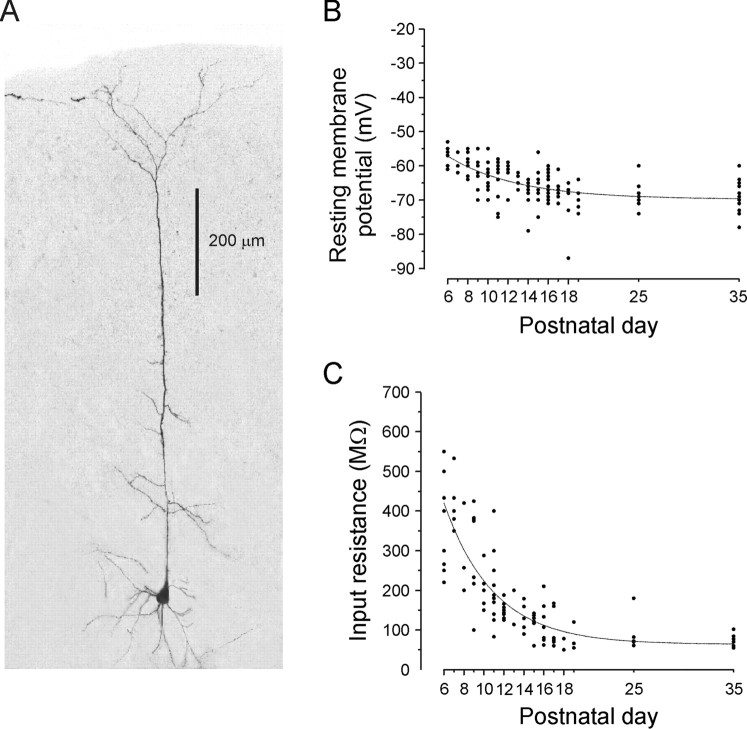
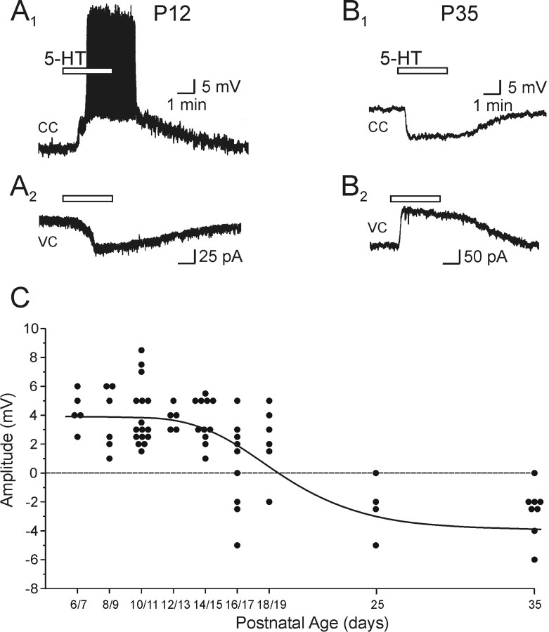
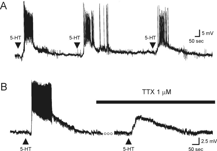
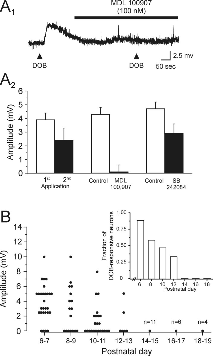
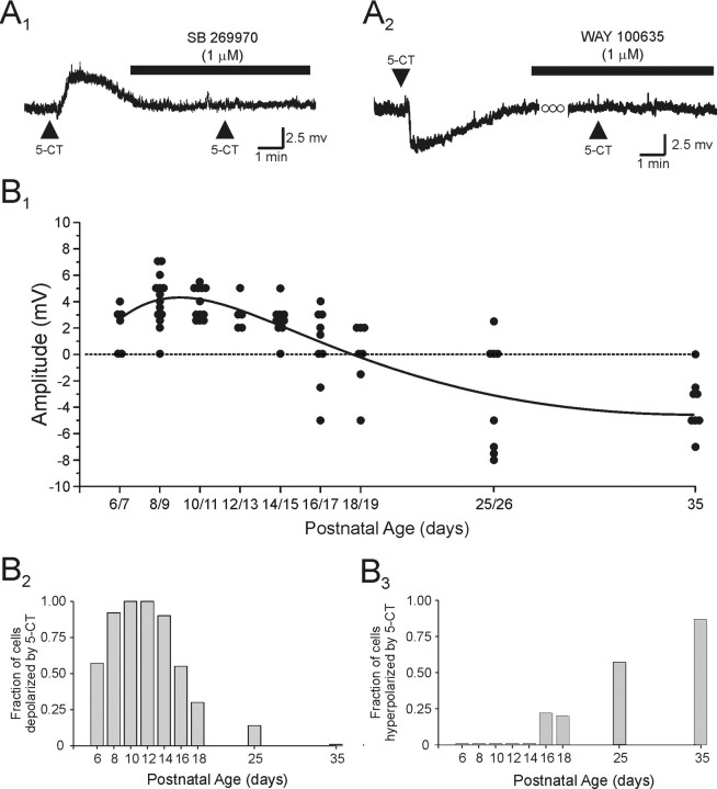
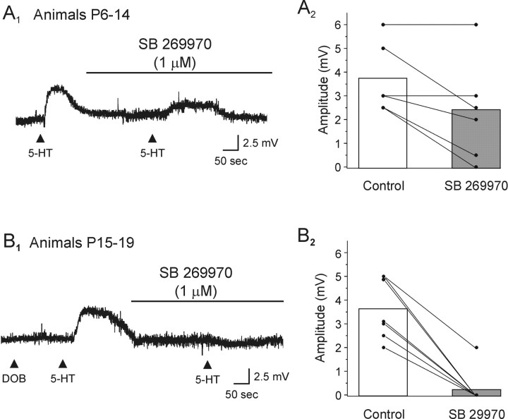
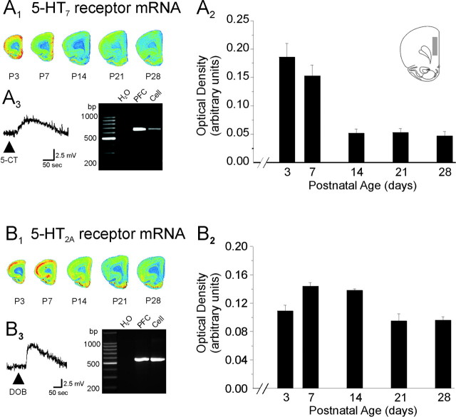
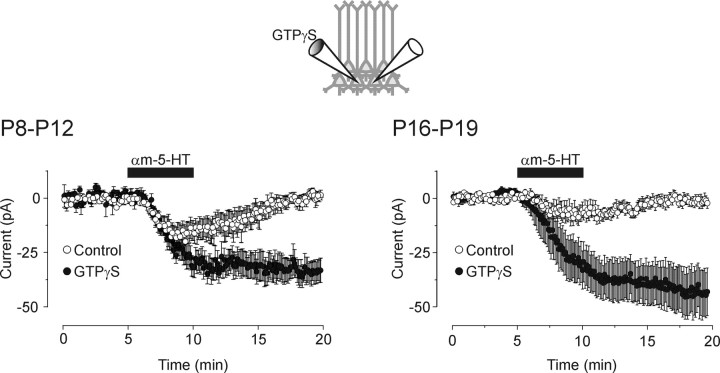
The developing prefrontal cortex receives a dense serotonergic innervation, yet little is known about the actions of serotonin [5-Hydroxytryptamine (5-HT)] in this region during development. Here, we examined the developmental regulation of 5-HT receptors controlling the excitability of pyramidal neurons of this region. Using whole-cell recordings in in vitro brain slices, we identified a dramatic shift in the effects of 5-HT on membrane potential during the postnatal developmental period. In slices derived from young animals [postnatal day (P) 6 to P19], administration of 5-HT elicits a robust depolarization of layer V pyramidal neurons, which gradually shifts to a hyperpolarization commencing during the third postnatal week. This progression is the result of coordinated changes in the function of 5-HT7 and 5-HT2A receptors, which mediate different aspects of the depolarization, and of 5-HT1A receptors, which signal the late developing hyperpolarization. The loss of the 5-HT7 receptor-mediated depolarization and the appearance of the 5-HT1A receptor-mediated hyperpolarization appears to reflect changes in receptor expression. In contrast, the decline in the 5-HT2A receptor depolarization with increasing age was associated with changes in the effectiveness with which these receptors could elicit a membrane depolarization, rather than loss of the receptors per se. Together, these results outline coordinated changes in the serotonergic regulation of cortical excitability at a time of extensive synaptic development and thus suggest a key role for these receptor subtypes in the postnatal development of the prefrontal cortex.


Figures

Figure 1.
Figure 1.
Electrophysiological properties of layer V pyramidal neurons of developing medial prefrontal cortex. A, Morphological reconstruction of one of the pyramidal neurons included in the present study. This neuron from a P12 animal was filled with Alexa 488 and reconstructed from a Z-stack using laser scanning confocal microscopy. B, Resting membrane potential of layer V pyramidal neurons across postnatal development. Note the tendency of these neurons to display more hyperpolarized membrane potentials with increasing age. C, Input resistance of layer V pyramidal neurons across postnatal development. The input resistance of these cells, determined using short hyperpolarizing current pulses in current clamp, markedly decreased during the first 2 weeks of postnatal life.
Figure 2.
Figure 2.
The effects of 5-HT on membrane excitability of layer V pyramidal neuron are regulated across postnatal development of the rat prefrontal cortex. A1, In a P12 animal, bath administration of 5-HT (30 μm) induced a prominent depolarization of sufficient magnitude to elicit action potential discharge in this layer V pyramidal neuron, as revealed in this current-clamp (CC) recording. The resting membrane potential (Vm) of this neuron was –64 mV. A2, The underlying inward current induced by 5-HT is shown in this current trace obtained in voltage-clamp (VC) mode from the same neuron. B1, In a P35 animal, bath administration of 5-HT (30 μm) induced a membrane hyperpolarization (Vm, –65 mV) in a layer V pyramidal neuron. B2, The underlying outward current is shown in this current trace obtained in voltage-clamp mode from the same neuron. C, Scatter graph showing the peak amplitude of the change in membrane potential of pyramidal neurons induced by drop application of 5-HT in the presence of TTX (1 μm). Data were collected from animals of all ages across the P6–P19 developmental window, binned at 2 d, and of P25 and P35 animals.
Figure 3.
Figure 3.
During the P6–P19 developmental period, 5-HT induces a robust, generally non-desensitizing membrane depolarization of layer V pyramidal neurons. A, Successive drop applications of 5-HT reliably depolarized and induced action potential discharge in this layer V pyramidal neuron from a P6 rat, as shown in this voltage trace (Vm, –66 mV). In this and subsequent figures, the arrow represents the time of agonist application. B, In this neuron from a P9 animal (Vm, –65 mV), application of 5-HT induced a membrane depolarization of sufficient magnitude to induce action potential discharge. TTX (1 μm) completely blocked the 5-HT-induced discharge of action potentials, revealing the underlying membrane depolarization. The open circles represent a period of 4 min.
Figure 4.
Figure 4.
Activation of 5-HT2A receptors depolarizes layer V pyramidal neurons of the rat prefrontal cortex. A1, Application of the selective 5-HT2 receptor agonist DOB induced a depolarization of this layer V pyramidal neuron (P11; Vm, –69 mV). This effect of DOB was blocked by the selective 5-HT2A receptor antagonist MDL 100907 (100 nm). This recording was conducted in the presence of 1 μm TTX. A2, Graph summarizing our pharmacological analysis of the DOB-induced depolarization. The response induced by DOB exhibited a small desensitization determined by two successive applications (8–12 min a part). The depolarizing response to DOB was completely blocked by bath administration of the selective 5-HT2A receptor MDL 100907 (100 nm), whereas the reduction of the DOB-induced depolarization by the selective 5-HT2C antagonist SB 242084 (100 nm) was similar to that expected from desensitization alone. B, Scatter graph showing the peak amplitude of the depolarization induced by application of DOB during the P6–P19 developmental period. Inset, Histogram showing the fraction of neurons that exhibited a depolarizing response to application of DOB across the same developmental window.
Figure 5.
Figure 5.
Activation of 5-HT7 receptors induces a membrane depolarization, which is gradually replaced by a 5-HT1A receptor-mediated hyperpolarization with increasing age. A1, In this neuron from a P10 animal, application of the 5-HT1/7 receptor agonist 5-CT induced a depolarization that was completely blocked by bath application of the selective 5-HT7 receptor antagonist SB 269970 (1 μm). This recording was conducted in the presence of 1 μm TTX. Vm, –70 mV. A2, In this neuron from a P25 animal (Vm, –68 mV), application of 5-CT induced a hyperpolarization, which was blocked by bath application of the selective 5-HT1A receptor antagonist WAY 100 635 (1 μm). B1, Scatter graph showing the peak change in membrane potential induced by application of 5-CT across postnatal development. B2, Histogram showing the fraction of neurons tested that exhibited a membrane depolarization in response to application of 5-CT as a function of the age of the animal. B2, Histogram showing the fraction of neurons that exhibited a membrane hyperpolarization in response to application of 5-CT as a function of the age of the animal.
Figure 6.
Figure 6.
The 5-HT-induced depolarization is mediated by activation of both 5-HT2A and 5-HT7 receptors. A1, In this neuron from a P10 animal, the depolarization induced by the application of 5-HT was only partially blocked by SB 269970 (1 μm). Vm, –65 mV. A2, Plot illustrating the effects of SB 269970 on the 5-HT-induced depolarization in slices derived from animals P6–P14. SB 269970 only partially antagonized the depolarization elicited by 5-HT in these slices.B1, In this DOB-unresponsive neuron taken from a P17 animal, the depolarization induced by 5-HT was completely blocked by SB 269970 (1 μm). Vm, –71 mV. B2, Plot illustrating the effects of SB 269970 on slices derived from animals P15–P19. SB 269970 (1 μm) essentially abolished the ability of 5-HT to depolarize pyramidal cells in these slices.
Figure 7.
Figure 7.
Expression of 5-HT2A and 5-HT7 receptors mRNA during the postnatal period. A1, B1, Pseudo-color autoradiographic image of cortical slices derived from rats of different ages are shown after hybridization with probes for the 5-HT7 receptor mRNA (A1) and 5-HT2A receptor mRNA (B1). The quantification of 5-HT7 (A2) and 5-HT2A (B2) receptor mRNA expression in the cingulate and prelimbic subdivisions of the medial prefrontal cortex is plotted across postnatal development. The area used to obtain the optical readings is illustrated in the inset of A2. This area corresponds to the area from which the electrophysiological recordings were performed. Graphed data depict optical density measurements expressed in arbitrary units. Data represent the mean ± SEM of four animals per age group. A3, Ethidium bromide-stained agarose gel showing the products of RT-PCR reactions amplifying 5-HT7 receptor mRNA from whole prefrontal cortex and from the pyramidal cell of which the voltage response to 5-CT is shown. Vm, –64 mV; P15 animal. The H2O lane depicts the results of a negative control using water as template. B3, Ethidium bromide-stained agarose gel showing the products of RT-PCR reactions amplifying 5-HT2A receptor mRNA from whole prefrontal cortex and from the pyramidal cell of which the voltage response to DOB is depicted. Vm, –71 mV; P10 animal.
Figure 8.
Figure 8.
Intracellular GTPγS facilitates the ability of 5-HT2A receptor to elicit an inward current in the developing prefrontal cortex. Left, Ensemble average of voltage-clamp recordings from P8–P12 slices showing that bath administration of αM-5-HT (10 μm) induced a small inward current in control cells (open circles). αM-5-HT (10 μm) induced a larger, nonrecovering, inward current in cells recorded with an intracellular recording solution supplemented with GTPγS (100 μm). Voltage-clamp recordings were simultaneously obtained from two close neighboring cells (as illustrated in the inset) with electrodes containing GTP alone or GTP plus GTPγS. Neurons were held at –70 mV, and the holding currents were sampled every 6 sec and then averaged across recordings. This plot is constructed from data obtained in six paired recordings. Right, Ensemble average of recordings obtained in P16–P19 slices showing that administration of αM-5-HT (10 μm) induced an inward current of much greater amplitude in cells perfused with intracellular GTPγS than in cells recorded with a control intracellular solution (n = 13 pairs). Note that the potentiation induced by GTPγS was itself of greater magnitude in slices derived from the older animals (P16–P19) than in those derived from younger animals (P8–P12).

References

    1. Adham N, Zgombick JM, Bard J, Branchek TA (1998) Functional characterization of the recombinant human 5-hydroxytryptamine7(a) receptor isoform coupled to adenylate cyclase stimulation. J Pharmacol Exp Ther 287: 508–514. - PubMed
    1. Aghajanian GK, Marek GJ (1999) Serotonin, via 5-HT2A receptors, increases EPSCs in layer V pyramidal cells of prefrontal cortex by an asynchronous mode of glutamate release. Brain Res 825: 161–171. - PubMed
    1. Andrade R, Chaput Y (1991) 5-Hydroxytryptamine4-like receptors mediate the slow excitatory response to serotonin in the rat hippocampus. J Pharmacol Exp Ther 257: 930–937. - PubMed
    1. Andrade R, Nicoll RA (1987) Pharmacologically distinct actions of serotonin on single pyramidal neurones of the rat hippocampus recorded in vitro J Physiol (Lond) 394: 99–124. - PMC - PubMed
    1. Andrade R, Malenka RC, Nicoll RA (1986) A G protein couples serotonin and GABAB receptors to the same channels in hippocampus. Science 234: 1261–1265. - PubMed

Publication types

MeSH terms

Substances

LinkOut - more resources