. 2004 May 19;24(20):4849-58.
doi: 10.1523/JNEUROSCI.0123-04.2004.

Efficient T-cell surveillance of the CNS requires expression of the CXC chemokine receptor 3

Affiliations

Efficient T-cell surveillance of the CNS requires expression of the CXC chemokine receptor 3

Jeanette Erbo Christensen et al. J Neurosci. .

Abstract

T-cells play an important role in controlling viral infections inside the CNS. To study the role of the chemokine receptor CXCR3 in the migration and positioning of virus-specific effector T-cells within the brain, CXCR3-deficient mice were infected intracerebrally with lymphocytic choriomeningitis virus (LCMV). Analysis of the induction phase of the antiviral CD8+ T-cell response did not reveal any immune defects in CXCR3-deficient mice. Yet, when mice were challenged with LCMV intracerebrally, most CXCR3-deficient mice survived the infection, whereas wild-type mice invariably died from CD8+ T-cell-mediated immunopathology. Quantitative analysis of the cellular infiltrate in CSF of infected mice revealed modest, if any, decrease in the number of mononuclear cells recruited to the meninges in the absence of CXCR3. However, immunohistological analysis disclosed a striking impairment of CD8+ T-cells from CXCR3-deficient mice to migrate from the meninges into the outer layers of the brain parenchyma despite similar localization of virus-infected target cells. Reconstitution of CXCR3-deficient mice with wild-type CD8+ T-cells completely restored susceptibility to LCMV-induced meningitis. Thus, taken together, our results strongly point to a critical role for CXCR3 in the positioning of effector T-cells at sites of viral inflammation in the brain.


Figures
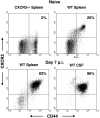
Figure 1.
Figure 1.
CXCR3 expression on CD8+ T-cells as a function of their activation state. Splenocytes were taken from uninfected WT and CXCR3-deficient mice. In addition, splenocytes and CSF cells were harvested from WT mice infected intracerebrally with 103 LD50 of LCMV 7 d earlier. All cells were stained with anti-CD8, anti-CD44, and anti-CXCR3. Gates were set for CD8+ cells; results are representative of more than five mice per group.
Figure 2.
Figure 2.
Lack of CXCR3 expression protects mice against a fatal outcome of LCMV-induced T-cell-mediated meningitis. WT and CXCR3-deficient mice were infected intracerebrally with 103 LD50 of LCMV, and mortality was registered (n = 23–31/group). Statistical evaluation was performed using the Mantel–Cox test: p < 0.0001. Brain virus titers of mice from parallel groups were determined and expressed as plaque-forming units per gram of organ (n = 3–4 mice/group).
Figure 3.
Figure 3.
Unimpaired CD8+ T-cell expansion in LCMV-infected CXCR3-deficient mice. Mice were infected intracerebrally with 103 LD50 of LCMV, and on 5–7 dpi, splenocytes were stained with anti-CD8, anti-VLA-4, and anti-L-selectin to determine the frequency of CD8+ T-cells with an activated phenotype. Gates were set for CD8+ T-cells, and the total number of VLA-4high/l-sellow cells/spleen is presented. To determine the frequency of virus-specific CD8+ T-cells, cells were stained for intracellular IFN-γ after in vitro stimulation with LCMV gp33-41 peptide for 5 hr. Gates were set for CD8+ T-cells, and the total number of VLA-4high/IFN-γ+ cells/spleen is presented. Averages ± SDs of four to eight mice per group are depicted.
Figure 4.
Figure 4.
Leukocyte recruitment to the CSF is not impaired in virus-infected CXCR3-deficient mice. A, Kinetics of leukocyte recruitment to the CSF of LCMV-infected mice. CXCR3-deficient and WT mice were infected intracerebrally with 103 LD50 of LCMV, and on the indicated days, CSF was harvested and the number of exudate cells in the CSF was determined; averages ± SD are depicted (n = 2–14 mice/group). B, Composition of the LCMV-induced cellular infiltrate as a function of time. CXCR3-deficient and WT mice were infected intracerebrally with 103 LD50, and on the indicated days, CSF was harvested and cells were stained with anti-CD8, anti-CD4, anti-Mac-1, and anti-B220 (B-cell marker) (n = 4–5/group).
Figure 5.
Figure 5.
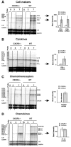
Comparison of cerebral mRNA expression after intracerebral LCMV infection. CXCR3-deficient and WT mice were infected intracerebrally with 103 LD50 of LCMV or injected with PBS (day 0 control). On the indicated days, total RNA was isolated from the brain of individual mice, and 20 μg was subjected to RPA analysis. Top lane, Expression of cell subset markers (A) and cytokines (B); bottom lane, expression of chemokines (C) and chemokine receptors (D).
Figure 6.
Figure 6.
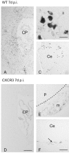
Failure of CD8+ T-cells to infiltrate the brain parenchyma in CXCR3-deficient mice. Immunohistochemistry analysis of brain sections from WT and CXCR3-deficient mice infected intracerebrally with 103 LD50 of LCMV. A–C, WT mice examined 7 dpi. A, Section taken from the level of the lateral choroid plexus (CP). CD8+ T-cells accumulate in brain regions near the ventricular system. B, CD8+ T-cells from the brain parenchyma shown at high magnification. C, T-cell accumulation in the cerebellum (Ce). CD8+ T-cells mainly occur in the white matter. D–F, CXCR3-deficient mice examined 7 dpi. D, Section taken at the level of the lateral ventricle containing the choroid plexus (CP). CD8+ T-cells do not accumulate in the brain parenchyma. E, Section from the ventral part of the brain stem showing accumulation of CD8+ T-cells in the meninges (m). The dashed line separates the meninges from the brain parenchyma (p), which is devoid of CD8+ T-cells. F, Section from the hind brain. CD8+ T-cells (arrow) localize to the meninges between the cerebellum (Ce) and the lower brain stem. Scale bars: A, D (in D), 200 μm; B, 10 μm; C, E, F (in F), 100 μm.
Figure 7.
Figure 7.
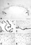
Immunohistochemical detection of LCMV virus in CXCR3-deficient (A–D) and WT (E) mice at 7 dpi. A, Section of the forebrain showing viral infection of choroid plexuses of the lateral and third ventricles (arrows) and inflammatory cells dispersed within the corpus callosum (open arrow). B, Labeling of choroid plexus epithelial cells shown at higher magnification. C, Section from the ventral part of the mesencephalon showing labeling of meninges. The subarachnoid space identified by an asterisk separates the inflamed meninges from unlabeled brain parenchyma. D, E, Sections of the corpus callosum of the CXCR3-deficient (D) and WT (E) mice. The number and distribution of inflammatory cells between the different mice are indistinguishable. Scale bars: A, 300 μm; B–E (in E), 100 μm.
Figure 8.
Figure 8.
CXCR3+ CD8+ T-cells restore susceptibility to LCMV-induced meningitis in CXCR3-deficient mice. CXCR3-deficient mice were infected intracerebrally with 103 LD50 of LCMV, and 3 d later, part of the mice were transfused with 3 × 10⁶ splenocytes from naive TCR transgenic mice expressing a TCR specific for LCMV gp33-41. Another group received the same number of transgenic splenocytes depleted of CD8+ T-cells (validated by flow cytometry). The mortality of these groups and a group of untransplanted virus-infected CXCR3-deficient mice was registered (n = 10–13 mice/group).
Figure 9.
Figure 9.
Unimpaired LCMV-induced T-cell-mediated inflammation in the footpad. LCMV-induced footpad swelling was assessed in mice infected locally in the right hind footpad with 200 pfu of LCMV. Virus-specific swelling was calculated as the difference in thickness of the infected right and the uninfected left hind foot. One of two similar experiments is presented; averages ± SD are depicted (n = 5 mice/group).

Similar articles

Cited by

References

    1. Andersson EC, Christensen JP, Marker O, Thomsen AR (1994) Changes in cell adhesion molecule expression on T cells associated with systemic virus infection. J Immunol 152: 1237–1245. - PubMed
    1. Andreasen SO, Christensen JP, Marker O, Thomsen AR (1999) Virus-induced non-specific signals cause cell cycle progression of primed CD8(+) T cells but do not induce cell differentiation. Int Immunol 11: 1463–1473. - PubMed
    1. Asensio VC, Campbell IL (1997) Chemokine gene expression in the brains of mice with lymphocytic choriomeningitis. J Virol 71: 7832–7840. - PMC - PubMed
    1. Asensio VC, Kincaid C, Campbell IL (1999) Chemokines and the inflammatory response to viral infection in the central nervous system with a focus on lymphocytic choriomeningitis virus. J Neurovirol 5: 65–75. - PubMed
    1. Balashov KE, Rottman JB, Weiner HL, Hancock WW (1999) CCR5(+) and CXCR3(+) T cells are increased in multiple sclerosis and their ligands MIP-1alpha and IP-10 are expressed in demyelinating brain lesions. Proc Natl Acad Sci USA 96: 6873–6878. - PMC - PubMed

Publication types

MeSH terms