Review
. 2004 Jun;13(6):1435-48.
doi: 10.1110/ps.03554604.

The ankyrin repeat as molecular architecture for protein recognition

Affiliations
Review

The ankyrin repeat as molecular architecture for protein recognition

Leila K Mosavi et al. Protein Sci. 2004 Jun.

Abstract

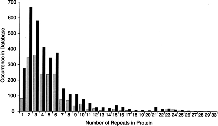
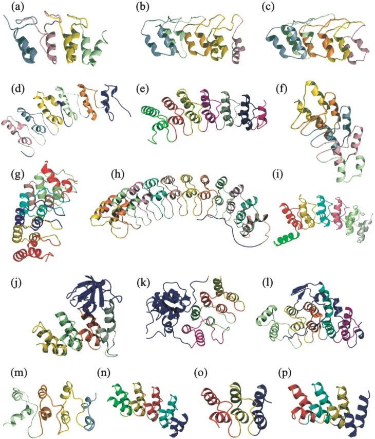
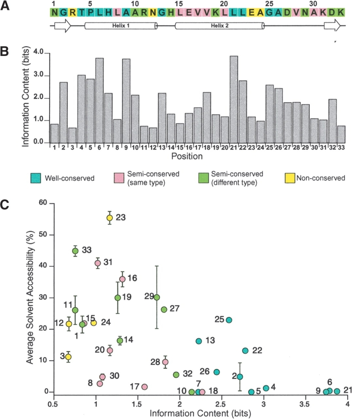
The ankyrin repeat is one of the most frequently observed amino acid motifs in protein databases. This protein-protein interaction module is involved in a diverse set of cellular functions, and consequently, defects in ankyrin repeat proteins have been found in a number of human diseases. Recent biophysical, crystallographic, and NMR studies have been used to measure the stability and define the various topological features of this motif in an effort to understand the structural basis of ankyrin repeat-mediated protein-protein interactions. Characterization of the folding and assembly pathways suggests that ankyrin repeat domains generally undergo a two-state folding transition despite their modular structure. Also, the large number of available sequences has allowed the ankyrin repeat to be used as a template for consensus-based protein design. Such projects have been successful in revealing positions responsible for structure and function in the ankyrin repeat as well as creating a potential universal scaffold for molecular recognition.
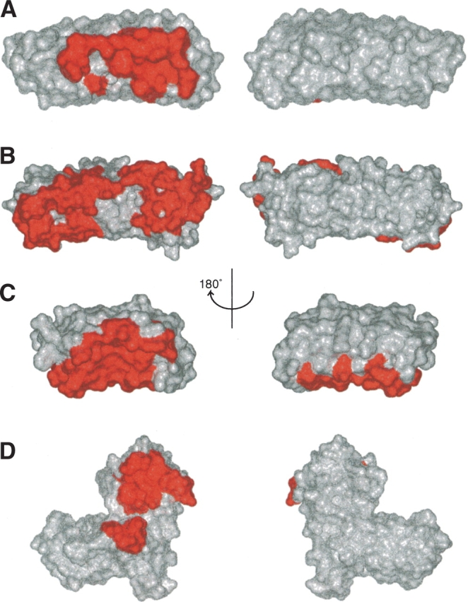

Figures

Figure 1.
Figure 1.
The distribution of the number of ankyrin repeats per protein (as of October 2003). Analysis was performed on the PFAM database, which contains 9689 ankyrin repeats (gray bars), and the SMART database, which contains 19,276 ankyrin repeats (black bars).
Figure 2.
Figure 2.
Ankyrin repeat proteins with high-resolution structures in the PDB. The ankyrin repeats have been colored differently to illustrate the packing interactions present in this fold. Cell cycle inhibitors (a) p16 (PDB ID: 1BI7), (b) p18 (1IHB), and (c) p19 (1AP7); (d) Iκ-Bα, inhibitor of Nf-κB (1NFI); (e) Bcl-3, a unique member of the Iκ-B family (1K1A); (f) Transcription factor GABP-β (1AWC); (g) Human oncoprotein Gankyrin (1UOH); (h) D34 region of human Ankyrin-R (1N11); (i) Ankyrin repeat domain of Drosophila signaling protein Notch (1OT8); (j) Tumor suppressor 53BP2 (1YCS); (k) Signaling protein PAP-β (1DCQ); (l) Transcriptional regulator Swi6 (1SW6); (m) Cardiomyogenic hormone myotrophin (2MYO). Designed ankyrin repeat proteins (n) Sank E3_5 (1MJ0), (o) 3ANK (1N0Q), and (p) 4ANK (1N0R).
Figure 3.
Figure 3.
Conservation level and information content in the ankyrin repeat. (A) The ankyrin repeat consensus sequence derived from statistical analysis of ~4400 ankyrin repeats used by Mosavi et al. 2002a, displaying the conservation level of each position as different colors. Corresponding secondary structure elements are displayed below. (B) The information content (bits) for each of the 33 positions in the ankyrin repeat calculated from the current version of PFAM database (containing ~9000 ankyrin repeat sequences) using the program ALPRO (Schneider and Stephens 1990). Higher information content implies that the residue is more conserved within the motif. (C) Comparison of the solvent accessibility of each position to the information content calculated by ALPRO. Average solvent accessibility was calculated for all 33 positions of the two internal repeats (repeat 2 and 3) in the structure of designed protein 4ANK (1N0R) with four identical repeats. Each marker is color coded according to the conservation-level classification of (Mosavi et al. 2002a) and labeled with the position number. Error bars represent the deviation from average for solvent accessibility.
Figure 4.
Figure 4.
(AF) Pairs of positions in the ankyrin repeat that have high covariation scores displayed on the structure of 4ANK (1N0R). Covariation was calculated as previously described using ~4400 ankyrin repeat sequences (Mosavi et al. 2002a). The backbone is displayed in gray, whereas the atoms of the residues shown are represented as blue spheres. All distances (red lines) are labeled.
Figure 5.
Figure 5.
Locations of the interaction interface in four different ankyrin repeat co-crystal structures. The residues involved in the interaction with protein partners are colored red in the surface representations. The β-hairpin/loop region is pointing directly towards reader (left) and away from the reader (right). (A) The interface between GABP-β and GABP-α from the co-crystal structure 1AWC, (B) the interface between Iκ-Bα and NFκ-B from the co-crystal structure 1NFI, (C) the interface between p16 and Cdk6 from the co-crystal structure 1BI7, (D) the interface between 53BP2 and p53 from the co-crystal structure 1YCS. Note that the lower red patch is the ankyrin repeat interface, while the upper patch is the SH3 domain interface.

Similar articles

Cited by

References

    1. Bae, J., Donigian, J.R., and Hsueh, A.J.W. 2003. Tankyrase 1 interacts with Mcl-1 proteins and inhibits their regulation of apoptosis. J. Biol. Chem. 278 5195–5204. - PubMed
    1. Batchelor, A.H., Piper, D.E., de la Brousse, F.C., McKnight, S.L., and Wolberger, C. 1998. The structure of GABPα/β: An ETS domain-ankyrin repeat heterodimer bound to DNA. Science 279 1037–1041. - PubMed
    1. Bateman, A., Birney, E., Cerruti, L., Durbin, R., Etwiller, L., Eddy, S.R., Griffiths-Jones, S., Howe, K.L., Marshall, M., and Sonnhammer, E.L. 2002. The Pfam protein families database. Nucleic Acids Res. 30 276–280. - PMC - PubMed
    1. Becker, T.M., Rizos, H., Kefford, R.F., and Mann, G.J. 2001. Functional impairment of melanoma-associated p16INK4a mutants in melanoma cells despite retention of cyclin-dependent kinase 4 binding. Clin. Cancer Res. 7 3282–3288. - PubMed
    1. Binz, H.K., Stumpp, M.T., Forrer, P., Amstutz, P., and Pluckthun, A. 2003. Designing repeat proteins: Well-expressed, soluble and stable proteins from combinatorial libraries of consensus ankyrin repeat proteins. J. Mol. Biol. 332 489–503. - PubMed

Publication types

LinkOut - more resources