

The structure of Ski8p, a protein regulating mRNA degradation: Implications for WD protein structure
- PMID: 15152089
- PMCID: PMC2279974
- DOI: 10.1110/ps.04704704
The structure of Ski8p, a protein regulating mRNA degradation: Implications for WD protein structure
Abstract
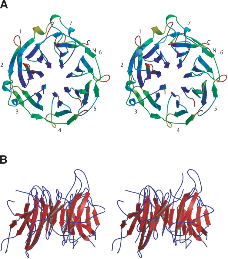
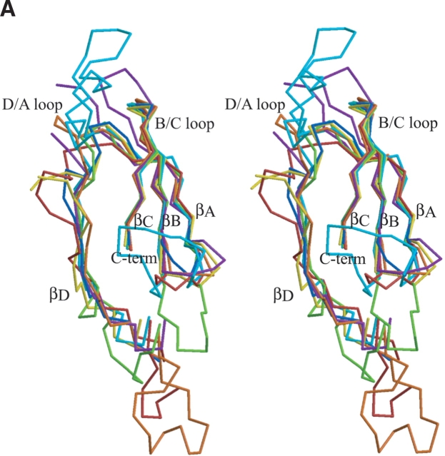
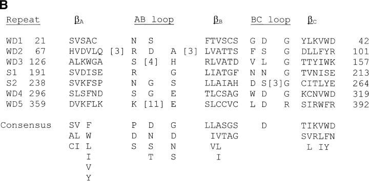
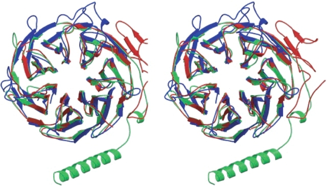
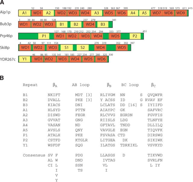
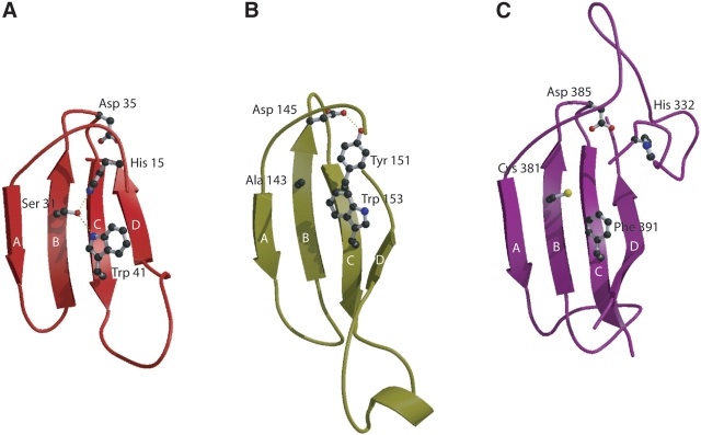
Ski8p is a 44-kD protein that primarily functions in the regulation of exosome-mediated, 3'--> 5' degradation of damaged mRNA. It does so by forming a complex with two partner proteins, Ski2p and Ski3p, which complete a complex that is capable of recruiting and activating the exosome/Ski7p complex that functions in RNA degradation. Ski8p also functions in meiotic recombination in complex with Spo11 in yeast. It is one of the many hundreds of primarily eukaryotic proteins containing tandem copies of WD repeats (also known as WD40 or beta-transducin repeats), which are short ~40 amino acid motifs, often terminating in a Trp-Asp dipeptide. Genomic analyses have demonstrated that WD repeats are found in 1%-2% of proteins in a typical eukaryote, but are extremely rare in prokaryotes. Almost all structurally characterized WD-repeat proteins are composed of seven such repeats and fold into seven-bladed beta propellers. Ski8p was thought to contain five WD repeats on the basis of primary sequence analysis implying a five-bladed propeller. The 1.9 A crystal structure unexpectedly exhibits a seven-bladed propeller fold with seven structurally authentic WD repeats. Structure-based sequence alignments show additional sequence diversity in the two undetected repeats. This demonstrates that many WD repeats have not yet been identified in sequences and also raises the possibility that the seven-bladed propeller may be the predominant fold for this family of proteins.
Figures
Similar articles
-
Crystal structure of Ski8p, a WD-repeat protein with dual roles in mRNA metabolism and meiotic recombination.Protein Sci. 2004 Oct;13(10):2673-84. doi: 10.1110/ps.04856504. Epub 2004 Aug 31. Protein Sci. 2004. PMID: 15340168 Free PMC article.
-
The yeast antiviral proteins Ski2p, Ski3p, and Ski8p exist as a complex in vivo.RNA. 2000 Mar;6(3):449-57. doi: 10.1017/s1355838200991787. RNA. 2000. PMID: 10744028 Free PMC article.
-
The 1.1-angstrom structure of the spindle checkpoint protein Bub3p reveals functional regions.J Biol Chem. 2005 Apr 8;280(14):13944-51. doi: 10.1074/jbc.M412919200. Epub 2005 Jan 11. J Biol Chem. 2005. PMID: 15644329
-
Phylogenetic, structural and functional relationships between WD- and Kelch-repeat proteins.Subcell Biochem. 2008;48:6-19. doi: 10.1007/978-0-387-09595-0_2. Subcell Biochem. 2008. PMID: 18925367 Review.
-
WD-repeat proteins: structure characteristics, biological function, and their involvement in human diseases.Cell Mol Life Sci. 2001 Dec;58(14):2085-97. doi: 10.1007/pl00000838. Cell Mol Life Sci. 2001. PMID: 11814058 Free PMC article. Review.
Cited by
-
The substrate specificity of eukaryotic cytosolic chaperonin CCT.Philos Trans R Soc Lond B Biol Sci. 2018 Jun 19;373(1749):20170192. doi: 10.1098/rstb.2017.0192. Philos Trans R Soc Lond B Biol Sci. 2018. PMID: 29735743 Free PMC article. Review.
-
Mechanism and Control of Meiotic DNA Double-Strand Break Formation in S. cerevisiae.Front Cell Dev Biol. 2021 Mar 2;9:642737. doi: 10.3389/fcell.2021.642737. eCollection 2021. Front Cell Dev Biol. 2021. PMID: 33748134 Free PMC article. Review.
-
Electrostatic and functional analysis of the seven-bladed WD beta-propellers.Evol Bioinform Online. 2008 Jun 13;4:203-16. doi: 10.4137/ebo.s743. Evol Bioinform Online. 2008. PMID: 19204818 Free PMC article.
-
Constitutive signal transduction by mutant Ssy5p and Ptr3p components of the SPS amino acid sensor system in Saccharomyces cerevisiae.Eukaryot Cell. 2005 Jun;4(6):1116-24. doi: 10.1128/EC.4.6.1116-1124.2005. Eukaryot Cell. 2005. PMID: 15947203 Free PMC article.
-
Three-dimensional structures of the flagellar dynein-microtubule complex by cryoelectron microscopy.J Cell Biol. 2007 Apr 23;177(2):243-52. doi: 10.1083/jcb.200609038. Epub 2007 Apr 16. J Cell Biol. 2007. PMID: 17438074 Free PMC article.
References
-
- Arora, C., Kee, K., Maleki, S., and Keeney, S. 2004. Antiviral protein Ski8 is a direct partner of Spo11 in meiotic DNA break formation, independent of its cytoplasmic role in RNA metabolism. Mol. Cell 13 549–559. - PubMed
-
- Branden, C.-I. 1991. The TIM barrel—the most frequently occurring folding motif in proteins. Curr. Opin. Stuct. Biol. 1 978–983.
Publication types
MeSH terms
Substances
Associated data
- Actions
Grants and funding
LinkOut - more resources
Full Text Sources
Molecular Biology Databases
