. 2004 Jun 16;23(12):2451-8.
doi: 10.1038/sj.emboj.7600246. Epub 2004 May 20.

A second human antiretroviral factor, APOBEC3F, is suppressed by the HIV-1 and HIV-2 Vif proteins

Affiliations

A second human antiretroviral factor, APOBEC3F, is suppressed by the HIV-1 and HIV-2 Vif proteins

Heather L Wiegand et al. EMBO J. .

Abstract

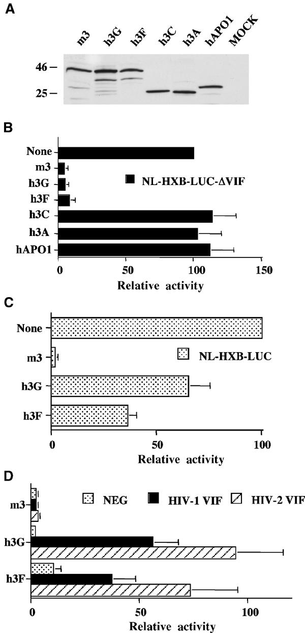
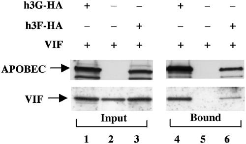
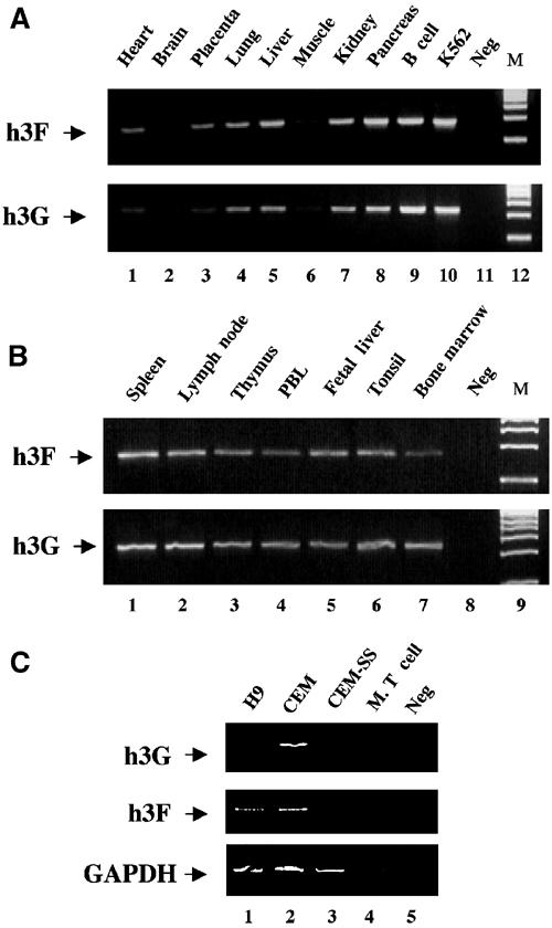
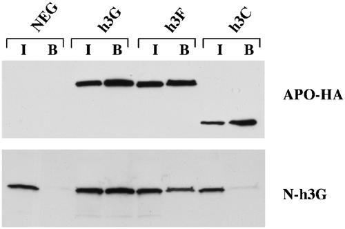
The HIV-1 Vif protein suppresses the inhibition of viral replication caused by the human antiretroviral factor APOBEC3G. As a result, HIV-1 mutants that do not express the Vif protein are replication incompetent in 'nonpermissive' cells, such as primary T cells and the T-cell line CEM, that express APOBEC3G. In contrast, Vif-defective HIV-1 replicates effectively in 'permissive' cell lines, such as a derivative of CEM termed CEM-SS, that do not express APOBEC3G. Here, we show that a second human protein, APOBEC3F, is also specifically packaged into HIV-1 virions and inhibits their infectivity. APOBEC3F binds the HIV-1 Vif protein specifically and Vif suppresses both the inhibition of virus infectivity caused by APOBEC3F and virion incorporation of APOBEC3F. Surprisingly, APOBEC3F and APOBEC3G are extensively coexpressed in nonpermissive human cells, including primary lymphocytes and the cell line CEM, where they form heterodimers. In contrast, both genes are quiescent in the permissive CEM derivative CEM-SS. Together, these data argue that HIV-1 Vif has evolved to suppress at least two distinct but related human antiretroviral DNA-editing enzymes.


Figures

Figure 1
Figure 1
Inhibition of productive HIV-1 infection by h3F is suppressed by both HIV-1 and HIV-2 Vif. (A) Western analysis of HA epitope-tagged forms of the indicated APOBEC proteins expressed in transfected 293T cells. (B) 293T cells were transfected with pNL-HXB-LUCΔVIF (1.5 μg) and the indicated APOBEC expression plasmid (125 ng). At 44 h after transfection, supernatant media were collected and used to infect CD4+ CXCR4+ 293T cells. A further 28 h later, these cells were lysed and induced luciferase activities were quantified. The average of three independent experiments with standard deviation is indicated. Activities are given relative to the virus obtained from the culture transfected with pNL-HXB-LUCΔVIF and the parental pcDNA3 plasmid, which was set at 100. (C) Similar to panel B, except that the Vif+ pNL-HXB-LUC indicator virus was used. (D) Similar to panel B, except that the cells were additionally transfected with 250 ng of a plasmid expressing HIV-1 Vif or HIV-2 Vif, or the pgΔVif plasmid as a negative control.
Figure 2
Figure 2
Specific packaging of h3F into HIV-1 virions is inhibited by both HIV-1 and HIV-2 Vif. 293T cells were transfected with 1.5 μg of the pNL4-3ΔVifΔEnv proviral expression plasmid, together with plasmids expressing HA-tagged forms of the indicated APOBEC proteins or the βarr2 control protein. Cells were also transfected with plasmids expressing HIV-1 or HIV-2 Vif, or a negative control plasmid (pgΔVif). At 44 h after transfection, supernatant media were harvested and released virus collected by ultracentrifugation, while the producer cells were collected and lysed. The cell and virion lysates were then subjected to gel electrophoresis, followed by Western analysis using a rabbit polyclonal antiserum specific for the HA tag or a monoclonal antibody specific for p24 Gag. While only the Gag Western performed with the disrupted virions is presented, closely similar results were also obtained using the cell lysates (data not shown). The level of expression of each APOBEC protein in the cell or virion lysates was quantified by scanning, and is presented for each panel normalized to the sample obtained in the absence of any Vif protein, which was set at 100. NA, not applicable.
Figure 3
Figure 3
The HIV-1 Vif protein specifically binds both h3G and h3F. 293T cells were transfected with an h3G or h3F expression plasmid, together with the HIV-1 Vif expression plasmid pgVif. The parental pcDNA3 plasmid served as a negative control. After 48 h, cells were lysed and a portion subjected to immunoprecipitation using an anti-HA monoclonal antibody. In all, 10% of the input lysate (lanes 1–3) and 25% of the bound fraction (lanes 4–6) were then subjected to electrophoresis, followed by Western analysis using rabbit polyclonal antisera specific for the HA epitope tag (upper panels) or the Vif protein (lower panels).
Figure 4
Figure 4
Analysis of the expression pattern of h3G and h3F mRNA by semiquantitative RT–PCR. (A) This experiment utilized a commercially obtained pre-normalized human multiple tissue cDNA panel. Positive controls represent non-normalized cDNA preparations derived from EBV-transformed B cells (lane 9) or human K562 cells (lane 10). Neg: no added cDNA. (B) Similar to panel A, except that a pre-normalized panel of human lymphoid tissue cDNAs was used. (C) This RT–PCR experiment utilized total RNA preparations derived from human H9, CEM or CEM-SS cells or mouse T cells. The GAPDH gene was used as an internal control. Neg: no added mRNA.
Figure 5
Figure 5
h3F and h3G form heteromultimers. This experiment was performed essentially as described in Figure 2A, except that cells were transfected with 500 ng each of N-tagged h3G and HA-tagged h3G, h3C or h3F (or pcDNA3 as a negative control). After immunoprecipitation using an HA-specific monoclonal, the recovered proteins were subjected to Western analysis using rabbit polyclonal antisera specific for the HA tag (panel A) or the N tag (panel B). Each panel shows both the input lysate (I, 10% of total) and the bound fraction (B, 25% of total).

Similar articles

Cited by

References

    1. Beale RCL, Petersen-Mahrt SK, Watt IN, Harris RS, Rada C, Neuberger MS (2004) Comparison of the differential context-dependence of DNA deamination by APOBEC enzymes: correlation with mutation spectra in vivo. J Mol Biol 337: 585–596 - PubMed
    1. Bogerd HP, Doehle BP, Wiegand HL, Cullen BR (2004) A single amino acid difference in the host APOBEC3G protein controls the primate species specificity of HIV-1 Vif. Proc Natl Acad Sci USA 101: 3770–3774 - PMC - PubMed
    1. Borman AM, Quillent C, Charneau P, Kean KM, Clavel F (1995) A highly defective HIV-1 group O provirus: evidence for the role of local sequence determinants in G → A hypermutation during negative-strand viral DNA synthesis. Virology 208: 601–609 - PubMed
    1. Chesebro B, Wehrly K, Nishio J, Perryman S (1992) Macrophage-tropic human immunodeficiency virus isolates from different patients exhibit unusual V3 envelope sequence homogeneity in comparison with T-cell-tropic isolates: definition of critical amino acids involved in cell tropism. J Virol 66: 6547–6554 - PMC - PubMed
    1. Conticello SG, Harris RS, Neuberger MS (2003) The Vif protein of HIV triggers degradation of the human antiretroviral DNA deaminase APOBEC3G. Curr Biol 13: 2009–2013 - PubMed

Publication types

Substances