. 2004;22(3):355-66.
doi: 10.1634/stemcells.22-3-355.

Characterization of putative stem cell phenotype in human limbal epithelia

Affiliations

Characterization of putative stem cell phenotype in human limbal epithelia

Zhuo Chen et al. Stem Cells. 2004.

Abstract

This study evaluated proposed molecular markers related to stem cell (SC) properties with the intention of characterizing a putative SC phenotype in human limbal epithelia. Human corneal and limbal tissues were cut in the vertical and horizontal meridians for histology, transmission electron microscopy (TEM), and immunostaining. Semiquantitative reverse transcriptase-polymerase chain reaction (RT-PCR) and in situ hybridization were used to evaluate gene expression. TEM showed that the limbal basal cells were small primitive cells. Immunostaining disclosed that p63, ABCG2 and integrin alpha9 were primarily expressed by the basal epithelial cells of limbus. Antibodies against integrin beta1, epidermal growth factor receptor (EGFR), K19, enolase-alpha, and CD71 stained the basal cells of the limbus more brightly than the suprabasal epithelia. Integrin alpha6, nestin, E-cadherin and connexin 43 did not stain the limbal basal cells, but the suprabasal epithelia of the cornea and limbus showed strong immunoreactivity. K3 and involucrin stained only corneal and limbal superficial cells. RT-PCR showed higher levels of p63, ABCG2 and integrin alpha9 mRNA, but lower levels of K3, K12 and connexin 43 expressed in the limbal epithelia than the corneal epithelia. In situ hybridization showed that p63 transcripts were located in basal layer of the limbal epithelium. This work suggests that the basal epithelial cells of the limbus are p63, ABCG2 and integrin alpha9 positive, and nestin, E-cadherin, connexin 43, involucrin, K3, and K12 negative, with relatively higher expression of integrin beta1, EGFR, K19, and enolase-alpha. This putative SC phenotype may facilitate the identification and isolation of limbal epithelial SCs.


Figures

Figure 1
Figure 1. Haematoxylin-eosin staining showing palisades of Vogt limbal architecture
A) A radial section through the central cornea showing about five layers of corneal epithelia and 8-10 layers of limbal epithelia. B) A tangential cross-section cut through the superior limbus showing the papilla-like epithelial columns and interspersed connective tissue of the palisades of Vogt environment.
Figure 2
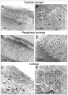
Figure 2. Transmission electron microscopic images of the central cornea (A, B), peripheral cornea (C, D) and limbus (E, F)
The basal cells (B) of central corneal epithelium are large columnar cells with low N/C ratio. The nucleus has loose chromatin and a pronounced nucleolus with coiled DNA, and the cytoplasm contains ribosomes and tonofilament. The basal cells (F) in limbal epithelia are smaller with a large N/C ratio, and they have barely detectable nucleolus with open DNA. The morphology of peripheral corneal epithelia is between central corneal and limbal epithelia (C, D). There is a Bowman's membrane (A, C) under the central and peripheral corneal basal epithelia with hemidesomosomes (B) or basal infoldings (D). The basal limbal epithelia (E) connect to basement membrane with invaginations, and the underneath stroma contains collagen and dilated capillaries. BM = Bowman's membrane; Cap = capillaries; Coil = coiled DNA; Col = collagen; Fold = basal infoldings; He = hemidesmosome; Inv = invaginations; N = nucleolus; Ri = ribosomes; To = tonofilaments.
Figure 3
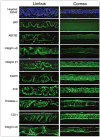
Figure 3. Immunofluorescent staining of proposed SC-associated markers, p63, ABCG2, integrin α9, integrin β1, EGFR, K19, enolase-α, CD71, and integrin α6 on frozen sections of limbus (left panels) and cornea (right panels)
Hoechst 33342 staining was used as counterstaining. Magnification: ×200.
Figure 4
Figure 4. Immunohistochemical staining for p63, ABCG2, and integrin α9 on limbal frozen sections
Only certain basal cells of limbal epithelia expressed nuclear p63, ABCG2, and integrin α9. Arrows indicate the positive stained cells.
Figure 5
Figure 5. Immunofluorescent staining for differentiation associated markers, K3, involucrin, connexin 43, E-cadherin, and nestin on frozen sections of limbus (left panels) and cornea (right panels)
Hoechst 33342 was used as a counterstaining. Magnification: ×200.
Figure 6
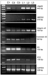
Figure 6. Semi-quantitative RT-PCR for SC-associated markers, p63 (440 bp), ABCG2 (379bp), and integrin α9 (123 bp), and differentiation associated markers, K3 (145bp), K12 (150 bp), and connexin 43 (154 bp) expressed by corneal (C1, C2, C3) and limbal (L1, L2, L3) epithelia from three fresh human corneal and limbal tissues
A 100 bp DNA ladder is shown in the first left lane. GAPDH, a housekeeping gene, was used as an internal control.
Figure 7
Figure 7. In situ hybridization of human limbal paraffin sections with [35S]-labeled p63 α riboprobes
The strong p63 signals were only located in the basal layer of limbal epithelia when hybridized with antisense p63 riboprobe (C, ×200 and D, ×400). There was no signal detected in control sections which were hybridized with sense riboprobe (A, ×200 and B, ×400).

References

    1. Hall PA, Watt FM. Stem cells: the generation and maintenance of cellular diversity. Development. 1989;106:619–633. - PubMed
    1. Potten CS, Loeffler M. Stem cells: attributes, cycles, spirals, pitfalls and uncertainties. Lessons for and from the crypt. Development. 1990;110:1001–1020. - PubMed
    1. Blau HM, Brazelton TR, Weimann JM. The evolving concept of a stem cell: entity or function? Cell. 2001;105:829–841. - PubMed
    1. Watt FM, Hogan BL. Out of Eden: stem cells and their niches. Science. 2000;287:1427–1430. - PubMed
    1. Lavker RM, Sun TT. Epidermal stem cells: properties, markers, and location. Proc Natl Acad Sci USA. 2000;97:13473–13475. - PMC - PubMed

Publication types

MeSH terms

Substances