

doi: 10.1128/AAC.48.6.2325-2330.2004.
Potential new anti-human immunodeficiency virus type 1 compounds depress virus replication in cultured human macrophages
Affiliations
- PMID: 15155246
- PMCID: PMC415615
- DOI: 10.1128/AAC.48.6.2325-2330.2004
Item in Clipboard
Potential new anti-human immunodeficiency virus type 1 compounds depress virus replication in cultured human macrophages
Antimicrob Agents Chemother.
2004 Jun.
Abstract
We report that the amiloride analogues 5-(N,N-hexamethylene)amiloride and 5-(N,N-dimethyl)amiloride inhibit, at micromolar concentrations, the replication of human immunodeficiency virus type 1 (HIV-1) in cultured human blood monocyte-derived macrophages. These compounds also inhibit the in vitro activities of the HIV-1 Vpu protein and might represent lead compounds for a new class of anti-HIV-1 drugs.
Figures
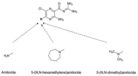
Chemical structures of amiloride, HMA, and DMA.
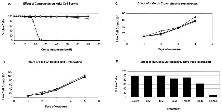
Effects of HMA on cell viability and proliferation. (A) HeLa cell monolayers were incubated in the presence of various concentrations of HMA (diamonds), DMA (squares), or amiloride (triangles) for 3 days. The cells were then detached from tissue culture wells by treatment with trypsin and were incubated with trypan blue to stain the dead cells. The proportion of live cells was determined by counting in a hemocytometer. (B and C) Effects of HMA at 0.1 μM (squares), 1.0 μM (triangles), and 10 μM (crosses) on the proliferation of CEMT4 cells (B) and T lymphocytes (C) cultured for 5 days. The ordinate shows the number of live cells after trypan blue staining. The diamonds indicate the proliferation of cells in drug-free control cultures. (D) Five-day-old MDMs (approximately 106 cells) were exposed to various concentrations of HMA for 7 days before they were harvested and stained with PI, followed by flow cytometry and fluorescence-activated cell sorter analysis. HMA was dissolved in dimethyl sulfoxide to give a concentration of 500 mM and was then serially diluted 1:9 in 0.1 N HCl and 1:4 in phosphate-buffered saline prior to final dilution in growth medium (RF10/10 [RPMI containing 10% human type AB serum and 10% fetal calf serum]). Solvent controls were prepared in the same way in which the compound was diluted. Ethanol (100%) was used as a positive control. The results are presented as the proportion of viable cells (PI negative) in HMA-treated cultures relative to the number of viable cells in the medium control.
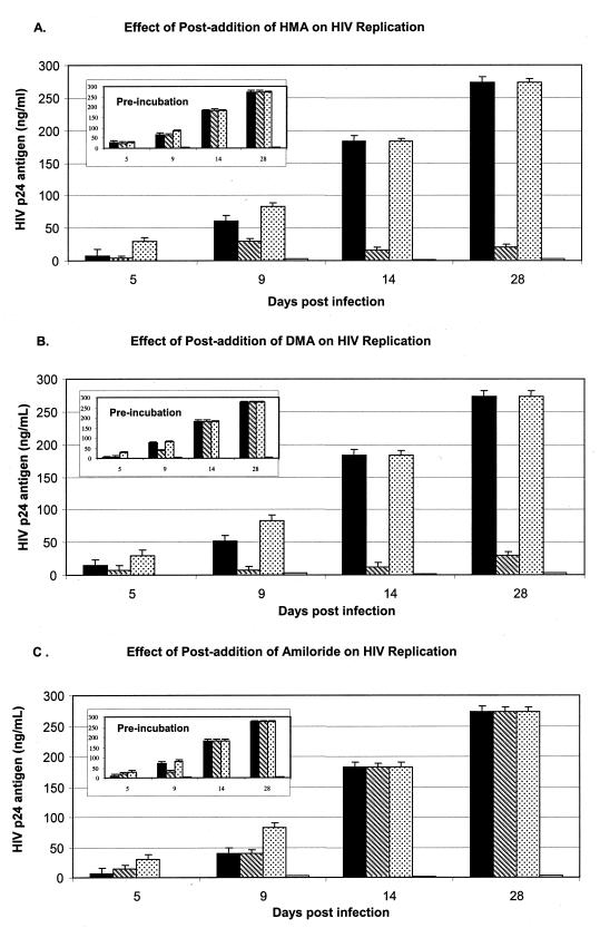
Effects of HMA (A), DMA (B), and amiloride (C) on replication of HIV-1BaL in MDMs. On day 1, 5-day-old macrophages were infected with HIV-1 at an MOI of 0.02/cell, and samples of the culture supernatant were taken at the indicated number of days postinfection for measurement of HIV-1 p24 antigen levels by ELISA. The groups of four bars for each day of measurement represent the average p24 levels for triplicate samples from cultures incubated in the presence of 1 μM HMA, 10 μM HMA, no-drug control, and 20 μM zidovudine, from left to right, respectively. The inset figures (labeled “preincubation”) show the effects of preincubation of identical cultures in the presence of the same concentrations of drug for 1 h prior to infection with HIV-1 and subsequent culture in drug-free medium. Replication of HIV-1 occurred at levels equivalent to those for the untreated controls, indicating that preexposure to compounds did not irreversibly inhibit an enzyme or damage a cellular structure necessary for HIV-1 replication or cell viability. The lower limit of p24 antigen detection is 7 pg/ml.
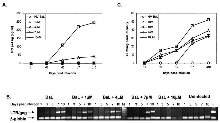
Effects of HMA on release of p24 antigen into MDM culture supernatants (A) and accumulation of intracellular HIV-1 DNA (B and C). Five-day-old MDM cultures were infected with HIV-1BaL (MOI, 0.02/cell) and exposed to 1, 4, 7, or 10 μM HMA for 10 days, as described in the text. Uninfected macrophages were used as negative controls, and cells infected with HIV-1BaL in the absence of HMA were considered positive controls. Either supernatants or cell lysates were collected at days 1, 3, 5, 7, and 10 postinfection. (A) Extracellular supernatants were assayed for p24 antigen by ELISA (Coulter). (B) Cell lysates were assayed for HIV DNA by a semiquantitative PCR with input cellular DNA. Simultaneously, a cellular 110-bp β-globin fragment was amplified by PCR. (C) The 320-bp LTR/gag band density, measured by densitometry with a Fluor-S Multi-Imager (gel documentation system; Bio-Rad), is plotted versus the number of days postinfection. By densitometry (data not shown) the β-globin band intensities are close to the control levels in all samples except those exposed to 10 μM HMA on days 7 and 10, in which a decrease of intensity of about 70% was measured. Potentially, this might indicate an increased rate of cell death for cells from this donor in the 10 μM HMA culture. Note, however, that this effect was not seen with HMA at concentrations of 7 μM or lower.
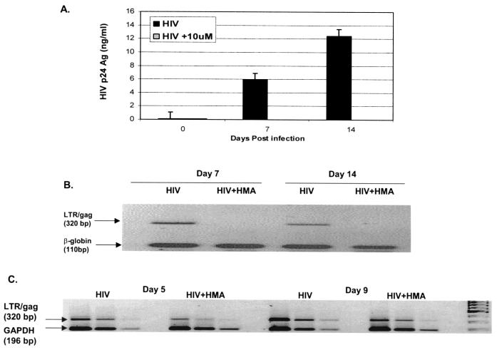
Effects of 10 μM HMA on production of p24 antigen (A), HIV-1 DNA (B), and HIV-1 RNA (C) in macrophage cultures. Macrophage cultures (in triplicate) were infected with HIV-1 and exposed to 10 μM HMA or the no-drug control over 14 days. (A) The levels of p24 antigen in culture supernatants, sampled on the indicated days postinfection, were measured by ELISA. As indicated, the solid bars for each day represent the results for the no-drug control and the shaded bars represent the presence of 10 μM HMA. (B) In the same experiment whose results are shown in panel A, total DNA was isolated from duplicate no-drug control and drug-treated macrophages at days 7 and 14 postinfection. PCRs were performed to simultaneously amplify a 320-bp LTR/gag fragment from HIV-1 DNA and a 110-bp β-globin gene fragment from cellular genomic DNA, and samples from the reaction mixtures were run on agarose gels and stained with ethidium bromide. Note that in contrast to the experiment whose results are shown in Fig. 4, with the cells from the donor whose results are shown here, the β-globin band intensity remains near the control level even after 14 days; this is consistent with observations that 10 μM HMA is well tolerated by MDMs from most donors. (C) Total RNA was isolated from control and drug-treated HIV-1-infected macrophages at day 5 and day 9 postinfection. RT-PCRs were performed to simultaneously amplify the 320-bp LTR/gag fragment from HIV-1 RNA and a 196-bp fragment from mRNA for the cellular housekeeping enzyme GAPDH. The groups of three lanes represent serial dilutions of the samples, used to facilitate quantitation by densitometry.
Similar articles
-
Amiloride derivatives block ion channel activity and enhancement of virus-like particle budding caused by HIV-1 protein Vpu.Eur Biophys J. 2002 Mar;31(1):26-35. doi: 10.1007/s002490100177. Eur Biophys J. 2002. PMID: 12046895
-
Antiviral efficacy of the novel compound BIT225 against HIV-1 release from human macrophages.Antimicrob Agents Chemother. 2010 Feb;54(2):835-45. doi: 10.1128/AAC.01308-09. Epub 2009 Dec 7. Antimicrob Agents Chemother. 2010. PMID: 19995924 Free PMC article.
-
Lack of effect of recombinant human growth hormone on the in vitro activities of antiretroviral drugs against human immunodeficiency virus type 1.Antimicrob Agents Chemother. 2004 Jun;48(6):2337-40. doi: 10.1128/AAC.48.6.2337-2340.2004. Antimicrob Agents Chemother. 2004. PMID: 15155249 Free PMC article.
-
Inhibition of HIV-1 replication by targeting the Rev protein.Leukemia. 1997 Apr;11 Suppl 3:134-7. Leukemia. 1997. PMID: 9209321 Review.
-
Multiple effects of interferon on the replication of human immunodeficiency virus type 1.Antiviral Res. 1994 Jul;24(2-3):205-19. doi: 10.1016/0166-3542(94)90068-x. Antiviral Res. 1994. PMID: 7526792 Review.
Cited by
-
Viral targets of acylguanidines.Drug Discov Today. 2012 Sep;17(17-18):1039-43. doi: 10.1016/j.drudis.2012.05.002. Epub 2012 May 8. Drug Discov Today. 2012. PMID: 22580299 Free PMC article. Review.
-
Novel Compound Inhibitors of HIV-1NL4-3 Vpu.Viruses. 2022 Apr 15;14(4):817. doi: 10.3390/v14040817. Viruses. 2022. PMID: 35458546 Free PMC article.
-
Requirement of a functional ion channel for Sindbis virus glycoprotein transport, CPV-II formation, and efficient virus budding.PLoS Pathog. 2022 Oct 3;18(10):e1010892. doi: 10.1371/journal.ppat.1010892. eCollection 2022 Oct. PLoS Pathog. 2022. PMID: 36191050 Free PMC article.
-
Modulation of influenza virus replication by alteration of sodium ion transport and protein kinase C activity.Antiviral Res. 2008 Nov;80(2):124-34. doi: 10.1016/j.antiviral.2008.05.008. Epub 2008 Jun 13. Antiviral Res. 2008. PMID: 18585796 Free PMC article.
-
The HIV-1 Vpu viroporin inhibitor BIT225 does not affect Vpu-mediated tetherin antagonism.PLoS One. 2011;6(11):e27660. doi: 10.1371/journal.pone.0027660. Epub 2011 Nov 14. PLoS One. 2011. PMID: 22110710 Free PMC article.
References
-
- Balliet, J. W., D. L. Kolson, G. Eiger, F. M. Kim, K. A. McGann, A. Srinivasan, and R. Collman. 1994. Distinct effects in primary macrophages and lymphocytes of the human immunodeficiency virus type 1 accessory genes vpr, vpu, and nef: mutational analysis of a primary HIV-1 isolate. Virology 200:623-631. - PubMed
-
- Carr, J. M., H. Hocking, P. Li, and C. J. Burrell. 1999. Rapid and efficient cell to cell transmission of human immunodeficiency virus infection from monocyte-derived macrophages to peripheral blood lymphocytes. Virology 265:319-329. - PubMed
-
- Du, B., A. Wolf, S. Lee, and E. Terwilliger. 1993. Changes in the host range and growth potential of an HIV-1 clone are conferred by the vpu gene. Virology 195:260-264. - PubMed
-
- Ewart, G. D., K. Mills, G. B. Cox, and P. W. Gage. 2002. Amiloride derivatives block ion channel activity and enhancement of virus-like particle budding caused by HIV-1 protein Vpu. Eur. Biophys. J. 31:26-35. - PubMed
Publication types
MeSH terms
Substances
LinkOut - more resources
Full Text Sources
Other Literature Sources
Medical
