. 2004 May;142(2):275-84.
doi: 10.1038/sj.bjp.0705786.

Molecular mechanisms of vasoselectivity of the 1,4-dihydropyridine lercanidipine

Affiliations

Molecular mechanisms of vasoselectivity of the 1,4-dihydropyridine lercanidipine

Susanne Wirtz et al. Br J Pharmacol. 2004 May.

Abstract

The effects of (S)- and (R)-lercanidipine on CHO cells stably expressing the cardiac (Ca(v)1.2a) or vascular (Ca(v)1.2b) splice variant of the L-type calcium channel pore subunit were studied, using whole-cell and single-channel patch-clamp measurements. Lercanidipine block of Ca(v)1.2b current was enantioselective. (S)-lercanidipine was 4.1-fold more potent. Experiments using acidic solutions (pH 6.8) revealed a 6.4-fold enhanced inhibitory effect of (S)-lercanidipine compared with physiological conditions (pH 7.4) indicating that the charged form mediates inhibition. At depolarised holding potential (-40 mV), (S)-lercanidipine exhibited a 35-fold greater potency, compared with standard conditions (-80 mV). A comparison of the concentration-dependent inhibition of Ca(v)1.2a with Ca(v)1.2b subunit currents by (S)-lercanidipine revealed only a 1.8-fold difference in IC(50), but the slope of the dose-response curve was much steeper (n(H)=2.3) with Ca(v)1.2a, compared with Ca(v)1.2b (n(H)=0.8). This indicates overlap between agonistic and antagonistic effects, predominant with the cardiac Ca(v)1.2a subunit. This idea is supported by transient stimulatory effects, and a slight leftward shift of the IV curves. These effects were more prominent for Ca(v)1.2a than for Ca(v)1.2b. Single-channel experiments confirmed typical features of calcium channel agonists such as prolonged channel openings, a component of lengthened openings, and an enhanced open probability in the presence of (S)-lercanidipine. Again, these findings were concentration-dependent and more pronounced for Ca(v)1.2a than for Ca(v)1.2b. Our data indicate a splice-variant predominant agonism as a new mechanism contributing to the vasoselectivity of lercanidipine, along with marked voltage-dependence of action.


Figures

Figure 1
Figure 1
(a) Time course of whole-cell peak current during a single experiment with CHO cells that stably express the vascular Cav1.2b pore subunit before and after addition of 10−7 M (S)-lercanidipine. Open symbols indicate control, filled symbols (S)-lercanidipine. The current was elicited from a holding potential of −80 mV, the test potential was +10 mV. The arrows indicate the time points of the traces presented in (b). (b) Original traces in the absence and presence of (S)-lercanidipine 10−7 M at the time points indicated by arrows in (a).
Figure 2
Figure 2
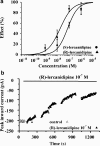
(a) Concentration–response curves for (S)- and (R)-lercanidipine obtained with CHO cells expressing the vascular Cav1.2b pore subunit of the L-type calcium channel. Each point represents data of six to eight experiments in the case of (S)-lercanidipine and of four to six experiments in the case of (R)-lercanidipine. The data were fitted by the Hill equation yielding an IC50 value of 1.8 × 10⁻⁸ M, Hill coefficient determined as nH=0.8 for (S)-lercanidipine and IC50=7.4 × 10⁻⁸ M, Hill coefficient fixed at nH=1 for (R)-lercanidipine. (b) Time course of whole-cell peak current during a single experiment with CHO cells that stably express the vascular Cav1.2b pore subunit before and after addition of 10−7 M (R)-lercanidipine. Open symbols indicate control, filled symbols (R)-lercanidipine. The current was elicited from a holding potential of −80 mV, the test potential was +10 mV.
Figure 3
Figure 3
Concentration–response curves for (S)-lercanidipine obtained in solutions buffered to pH 6.8 or 7.4 with CHO cells expressing the vascular Cav1.2b pore subunit of the L-type calcium channel. Each point represents means±s.e.m. of four to five experiments (pH 6.8) or six to eight experiments (pH 7.4). * Indicates P<0.05 (post-tests among identical concentrations). The data were fitted by the Hill equation yielding an IC50 value of 2.8 × 10⁻⁹ M, Hill coefficient fixed at nH=1 for pH 6.8 and IC50=1.8 × 10⁻⁸ M, Hill coefficient nH=0.8 for pH 7.4.
Figure 4
Figure 4
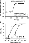
(a) Time course of whole-cell peak current during a single experiment with CHO cells that stably express the vascular Cav1.2b pore subunit before and after addition of 10−8 M (S)-lercanidipine at a holding potential (HP) of −40 mV. The test potential was +10 mV. Open symbols indicate control, filled symbols (S)-lercanidipine 10−8 M. (b) Concentration–response curves for (S)-lercanidipine obtained with CHO cells expressing the vascular Cav1.2b subunit of the L-type calcium channel at two different holding potentials of −40 and −80 mV. Values indicate means±s.e.m. of three to five experiments (HP −40 mV) and of six to eight experiments (HP −80 mV). * Indicates P<0.05 (post-tests among identical concentrations). IC50 determined by Hill analysis of the data is 5.2 × 10⁻¹⁰ M, Hill coefficient nH=0.9 for the depolarised holding potential −40 mV and IC50=1.8 × 10⁻⁸ M, Hill coefficient nH=0.8 for the holding potential −80 mV.
Figure 5
Figure 5
Concentration–response curves for (S)-lercanidipine obtained with CHO cells expressing the cardiac Cav1.2a subunit or the vascular Cav1.2b subunit of the L-type calcium channel. Values indicate means±s.e.m. of three to six experiments (Cav1.2a) and of six to eight experiments (Cav1.2b). IC50 determined by Hill equation is 3.3 × 10⁻⁸ M, Hill coefficient nH=2.3 for the cardiac Cav1.2a subunit and IC50=1.8 × 10⁻⁸ M, Hill coefficient nH=0.8 for the vascular Cav1.2b pore subunit.
Figure 6
Figure 6
A slight stimulatory effect of 10−8 M (S)-lercanidipine on the cardiac Cav1.2a subunit current was observed in three of nine experiments. (a) Time course of whole-cell peak current. Open symbols indicate control, filled symbols (S)-lercanidipine. The arrows indicate the time points of the traces presented in (b). (b) Original traces before and after addition of 10−8 M (S)-lercanidipine. (c) Current density–voltage relationship in the absence and presence of 10−8 M (S)-lercanidipine of the same experiment as in (a) and (b). Open symbols indicate control data, filled symbols data at time ∼300 s after (S)-lercanidipine application.
Figure 7
Figure 7
Effect of 10−7 M (S)-lercanidipine on the L-type calcium current through a single cardiac Cav1.2a pore subunit in a cell-attached patch (150 ms pulse length, holding potential −100 mV, test pulse +10 mV, 1.67 Hz). (a) Five consecutive traces before (left) and after application (right) of 10−7 M (S)-lercanidipine. Bottom rows: ensemble average current of all traces of each experiment. Scale bars: 20 ms/1 pA (single traces) and 20 ms/5 fA (ensemble average current). (b) Open time distribution before (left, open columns) and after 10−7 M (S)-lercanidipine (right, filled columns). (c) Time course of open probability before and after application of 10−7 M (S)-lercanidipine.
Figure 8
Figure 8
Effect of 10−5 M (S)-lercanidipine on the L-type calcium channel current obtained with a single CHO cell stably expressing the cardiac Cav1.2a pore subunit using the cell-attached configuration (100 ms pulse length, holding potential −100 mV, test pulse +10 mV, 1.67 Hz). (a) Open time distributions before (left, open columns) and after application of 10−5 M (S)-lercanidipine (right, filled columns). (b) Time course of open probability before and after application of 10−5 M (S)-lercanidipine.
Figure 9
Figure 9
Effects of (S)-lercanidipine 10−7 M on the L-type calcium channel activity in a cell-attached patch (150 ms pulse length, holding potential −100 mV, test pulse +10 mV, 1.67 Hz) obtained with a CHO cell stably expressing the vascular Cav1.2b pore subunit. (a) Open time distributions before (left, open columns) and after application of 10−7 M (S)-lercanidipine (right, filled columns). (b) Time course of open probability before and after application of 10−7 M (S)-lercanidipine.

Similar articles

Cited by

References

    1. ANGELICO P., GUARNERI A., LEONARDI A., TESTA R. Vascular-selective effect of lercanidipine and other 1,4-dihydropyridines in isolated rabbit tissues. J. Pharm. Pharmacol. 1999;51:709–714. - PubMed
    1. BAINDUR N., RUTLEDGE A., TRIGGLE D.J. A homologous series of permanently charged 1,4-dihydropyridines: novel probes designed to localize drug binding sites on ion channels. J. Med. Chem. 1993;36:3743–3745. - PubMed
    1. BANGALORE R., BAINDUR N., RUTLEDGE A., TRIGGLE D.J., KASS R.S. L-type calcium channels: asymmetrical intramembrane binding domain revealed by variable length, permanently charged 1,4-dihydropyridines. Mol. Pharmacol. 1994;46:660–666. - PubMed
    1. BEAN B.P. Nitrendipine block of cardiac calcium channels: high affinity binding to the inactivated state. Proc. Natl. Acad. Sci. U.S.A. 1984;81:6388–6392. - PMC - PubMed
    1. BECHEM M., HOFFMANN H. The molecular mode of action of the Ca agonist (−) Bay K 8644 on the cardiac Ca channel. Pflügers Arch. 1993;424:343–353. - PubMed

Publication types

LinkOut - more resources