. 2004 May 15;18(10):1209-21.
doi: 10.1101/gad.300104.

The T-box transcription factor Tbx18 maintains the separation of anterior and posterior somite compartments

Affiliations

The T-box transcription factor Tbx18 maintains the separation of anterior and posterior somite compartments

Markus Bussen et al. Genes Dev. .

Abstract

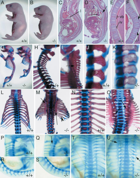
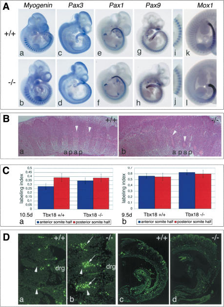
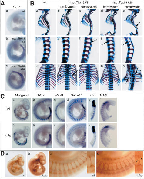
The compartmentalization of somites along their anterior-posterior (AP) axis is pivotal to the segmental organization of the vertebrate axial skeleton and the peripheral nervous system. Anterior and posterior somite halves contribute to different vertebral elements. They are also characterized by different proliferation rates and properties with respect to neural crest cell migration and spinal nerve passage. AP-somite polarity is generated in the anterior presomitic mesoderm by Mesp2 and Delta/Notch signaling. Here, we demonstrate that maintenance of AP-somite polarity is mediated by the T-box transcription factor Tbx18. Mice deficient for Tbx18 show expansion of pedicles with transverse processes and proximal ribs, elements derived from the posterior lateral sclerotome. AP-somite polarity is established in Tbx18 mutant embryos but is not maintained. During somite maturation, posterior somite compartments expand most likely because of posterior cells invading the anterior somite half. In the anterior lateral sclerotome, Tbx18 acts as an antiapoptotic factor. Ectopic expression experiments suggest that Tbx18 can promote anterior at the expense of posterior somite compartments. In summary, Tbx18 appears to act downstream of Mesp2 and Delta/Notch signaling to maintain the separation of anterior and posterior somite compartments.


Figures

Figure 1.
Figure 1.
Targeted disruption of the Tbx18 locus. (A) Schematic representation of the targeting strategy. Restriction map of the wild-type locus with boxes representing the first four exons of Tbx18; coding regions are shown in black, noncoding in white. Arrows show location and orientation of PCR primers. Fragments used as RFLP probes are shown. The EcoRV fragment designated as 3′ detects the EcoRI–RFLP shown in B. (A) ApaI; (N) NotI; (RI) EcoRI; (RV) EcoRV; (neo) loxP-flanked neomycin selection cassette; (IRES) internal ribosomal entry site. (B) Southern blot analysis of EcoRI-digested genomic DNA extracted from E18.5 embryos derived from intercrosses of Tbx18/+ mice. Genotypes are indicated above each lane. The 16-kb and 8.5-kb band represent the wild-type and the mutant allele, respectively. (C) PCR genotyping of embryos from heterozygous matings using primers specific for the wild-type (upper panel) and the mutant allele (lower panel), respectively. The genotypes are indicated above each lane.
Figure 2.
Figure 2.
Morphology and skeletal defects of newborns and embryos mutant for Tbx18.(A,B) External morphology. The Tbx18–/– pup (B) has a shorter body axis than the wild type (A). (BF) Hematoxylin-and-eosin-stained sagittal sections of newborn Tbx18–/– pups and wild-type littermates. The mutant (D) shows a massive kink in the vertebral column of the thoracic region (arrow); the lung (l) is not inflated. Higher magnification of the thoracic vertebral column showing regular spacing between vertebral bodies (vb) and intervertebral discs (iv) in the wild type (E), and distorted nuclei pulposi (arrows) within thinner intervertebral discs in the mutant (F). (GO) Skeletal preparations of Tbx18–/– newborns. Lateral view of whole skeletons (G) and higher magnification of the cervical/upper thoracic (H,I) and lumbar (J,K) regions of wild-type and mutant pups. (I) The arrow points to a small orifice for a spinal nerve within the pedicle band in the mutant. (K) Note altered shape of pedicles (p) in the mutant. (L,M) Dorsal views showing smaller rib cage with ossification band in the proximal region (arrows) and fusions of distal ribs (arrowhead) in Tbx18–/– pups (M). (N,O) Ventral view of a wild-type (N) and a Tbx18–/– (O) thoracic region showing that rib heads remain separate in the mutant (arrows). (PU) Alcian blue stainings of the cartilaginous preskeleton in whole E14.5 Tbx18–/– embryos. Lateral views of upper cervical (P,Q) and lumbar regions (R,S); dorsal views of rib cages (T,U) of wild type (P,R,T) and Tbx18–/– embryos (Q,S,U). Arrows point to expanded pedicles (Q) and proximal ribs (U) in mutant embryos.
Figure 3.
Figure 3.
Somite development and differentiation in Tbx18 mutant embryos. (A) In situ hybridization analysis for markers of somite differentiation at E10.5, in the wild-type (panels a,c,e,g,i,k) and in Tbx18–/– mutants (panels b,d,f,h,j,l). Markers for myotome (Myogenin, panel b), dermomyotome (Pax3, panel d), and ventral sclerotome (Pax1, panel f) are unchanged in Tbx18–/– embryos. Progressive loss of polarized Pax9 (panels g,h, and higher magnification of thoracic somites in panels i,j) and Mox1 (panels k,l) expression. (B) Histological analysis of sclerotome compartmentalization in cervical regions of Tbx18–/– embryos at E10.5. In the wild type (panel a), a regular pattern of less condensed anterior (a) and more condensed posterior (p) lateral sclerotomal cells is visible. (Panel b) In Tbx18–/– embryos, cell densities are similar in both compartments. Arrowheads point to spinal nerves passing through anterior halves. (C) Analysis of cell proliferation in somites of Tbx18–/– embryos. Labeling index is defined by the ratio of BrdU-positive cells to total cell number in the analyzed area. (Panel a) At E10.5d, labeling indices between anterior and posterior sclerotomal halves of cervical somites are clearly different in the wild type (anterior, 0.278 ± 0.036 vs. posterior, 0.386 ± 0.048). In Tbx18–/– embryos, cell proliferation in anterior somite halves as judged by the labeling index has almost reached the rate of posterior halves (anterior, 0.348 ± 0.051, vs. posterior, 0.383 ± 0.076). The increase of proliferation between anterior halves of wild type and mutant is statistically significant (p = 0.0022). (Panel b) In forming somites at E9.5, no difference in cell proliferation was detected between anterior and posterior halves of wild-type (anterior, 0.561 ± 0.091; posterior, 0.548 ± 0.094) and Tbx18–/– embryos (anterior, 0.0624 ± 0.041; posterior, 0.599 ± 0.062), respectively. The overall increase in the mutant is not statistically relevant. (D) Analysis of apoptosis in somites of Tbx18–/– embryos. (Panels a,b) E10.5. Increased apoptosis (arrows) in the anterior lateral sclerotome of cervical somites of Tbx18–/– embryos (panel b) compared with wild type (panel a). Arrowheads point to spinal nerves. (Panels c,d) E9.5d. No difference in apoptosis was detected in somites of Tbx18–/– embryos (panel d), respectively.
Figure 4.
Figure 4.
Regulation and function of Tbx18 in anterior–posterior somite polarity. (A) Whole-mount in situ hybridization analysis of Tbx18 expression in embryos with defects in somitogenesis at E10.5. (Panels a,c,e,f) Whole embryos. (Panels b,d,f,h) Higher magnification of posterior trunk regions. (Panels a,b) In the wild type, Tbx18 expression is restricted to anterior halves of prospective and definitive somites. (Panels c,d) Tbx18 is expressed throughout the anterior PSM and somites in Dll1–/– embryos. (Panels e,f) Expression boundaries are fuzzy in pudgy/pudgy (Dll3–/–) embryos. (Panels g,h) In Uncx4.1–/– embryos, there is continuous expression of Tbx18 in posterior somite halves but not in the PSM region (arrows). (B) Whole-mount in situ hybridization analysis of expression of markers for early AP-somite polarity in posterior trunks at E10.5. Markers as indicated in the figure and described in the text are not changed in Tbx18–/– (panels b,d,f,h,j,l,n,p) compared with wild-type (panels a,c,e,g,i,k,m,o) embryos. (E B2) EphrinB2. (C) In situ hybridization analysis of Uncx4.1 expression at E10.5 in Tbx18–/– embryos. Whole embryos (panels a,b), posterior trunk regions (panels c,d), and sections of cervical somite regions (panels e,f) with anterior to the right. (Panels a,c,e) In the wild type, Uncx4.1 expression is confined to posterior somite halves. (Panels b,d,f) In Tbx18–/– embryos, Uncx4.1 expression progressively expands into the anterior somite compartment. (D) Analysis of peripheral nervous system development in Tbx18–/– embryos. (Panels a,b) HE-stained parasagittal sections of cervical regions at E13.5. In the wild type (panel a), dorsal root ganglia (drg) are clearly separated; in Tbx18–/– embryos (panel b), they are fused. (Panels c–f) Antineurofilament staining to reveal the organization of spinal nerve projections at E10.5. In the wild type (panels c,e), spinal nerves traverse the anterior half of each mature somite, and in the mutant some spinal nerves are lacking (arrows in panel d); partly, the ones present appear thinner than in the wild type (arrows and arrowheads in panel f), particularly at their base.
Figure 5.
Figure 5.
Ectopic expression of Tbx18 in somites leads to defects in AP-somite polarity and lateral sclerotome development. (A) In situ hybridization analysis of expression of Tbx18-GFP-fusion transcripts in E10.5 transgenic embryos. Embryos hemizygous of msd::Tbx18 line #2 (panel b) and msd::Tbx18 line #20 (panel c) show transgene expression in the PSM, the somitic mesoderm, and the myotome. (B) Skeletal malformations in msd::tbx18 transgenic at E18.5. Genotypes of embryos are indicated above each row. (Panels a–e) Lateral views of whole skeletal preparations of wild-type and transgenic embryos. Arrow in panel e points to reduced pedicles along the vertebral column. (Panels f–j) Higher magnification of the lumbar region of the skeletal preparations shown in panels a–e. Arrows in panels h, i, and j point to reduced pedicles. (Panels k–o) Higher magnification of the rib cage in a dorsal view of the skeletal preparations shown in panels a–e; arrows in l, m, and o point to reduced or missing proximal ribs. (C) In situ hybridization analysis of somite differentiation and polarization in homozygotes of msd::Tbx18 (tg/tg) line #20 at E10.5. Genotypes are indicated in the figure. (Panel g) Myogenin expression is unchanged. (Panel h) Mox1 expression in posterior somite halves is reduced to a thin stripe in the interlimb region of transgenic embryos. (Panel i) The strong posterior Pax9 expression domain is reduced in the interlimb region of msd::Tbx18 transgenic embryos. Anterior is up. (Panel j) Uncx4.1 expression analysis reveals reduction of posterior somite compartments in transgenic embryos. Note that Uncx4.1 expression is normal in the most recently formed five to six somites. (Panel k) Dll1 expression in the PSM and in the most recently formed three somites is unchanged in transgenic embryos, then expression gets progressively weaker. (Panel l) EphrinB2 (E B2) expression is normal in the first four to five somites of transgenic embryos, then it gets weaker. (D) Antineurofilament staining to reveal the organization of spinal nerve projections in homozygotes of msd::Tbx18 line #20 at E10.5. The metameric organization of spinal nerve projections is maintained in transgenic embryos (panel b); however, they appear less bundled (panel d). Higher magnification of the interlimb region of the embryos shown in panels a and b.Anterioristothe left. (Panel d) Arrows in the small inset figure point to colaterals leaving the nerve.

References

    1. Aoyama H. and Asamoto, K. 2000. The developmental fate of the rostral/caudal half of a somite for vertebra and rib formation: Experimental confirmation of the resegmentation theory using chick-quail chimeras. Mech. Dev. 99: 71–82. - PubMed
    1. Barrantes del Barco I., Elia, A.J., Wünsch, K., Hrabe De Angelis, M., Mak, T.W., Rossant, R., Conlon, R.A., Gossler, A., and Pompa, J-L. 1999. Interaction between L-fringe and Notch signalling in the regulation of boundary formation and posterior identity in the presomitic mesoderm of the mouse. Curr. Biol. 9: 470–480. - PubMed
    1. Beckers J., Caron, A., Hrabe de Angelis, M., Hans, S., Campos-Ortega, J.A., and Gossler, A. 2000. Distinct regulatory elements direct Delta1 expression in the nervous system and paraxial mesoderm of transgenic mice. Mech. Dev. 95: 23–34. - PubMed
    1. Bettenhausen B., de Angelis, M.H., Simon, D., Guénet, J.-L., and Gossler, A. 1995. Transient and restricted expression during mouse embryogenesis of Dll1, a murine gene closely related to Drosophila Delta. Development 121: 2407–2418. - PubMed
    1. Biben C., Stanley, E., Fabri, L., Kotecha, S., Rhinn, M., Drink-water, C., Lah, M., Wang, C.C., Nash, A., Hilton, D., et al. 1998. Murine cerberus homologue mCer-1: A candidate anterior patterning molecule. Dev. Biol. 194: 135–151. - PubMed

Publication types