

Experimental pneumococcal meningitis: impaired clearance of bacteria from the blood due to increased apoptosis in the spleen in Bcl-2-deficient mice
- PMID: 15155612
- PMCID: PMC415656
- DOI: 10.1128/IAI.72.6.3113-3119.2004
Experimental pneumococcal meningitis: impaired clearance of bacteria from the blood due to increased apoptosis in the spleen in Bcl-2-deficient mice
Abstract
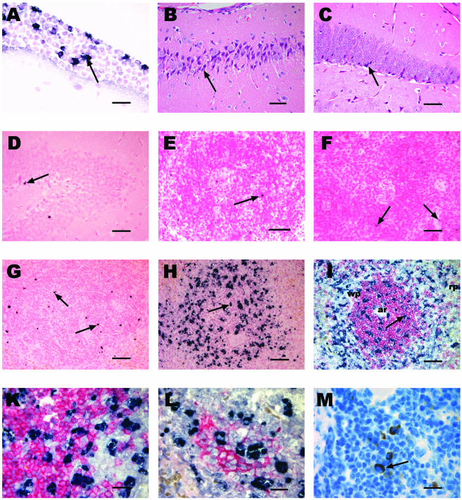
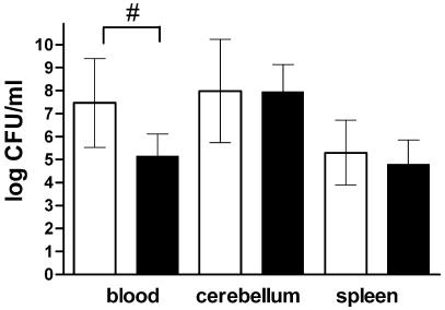
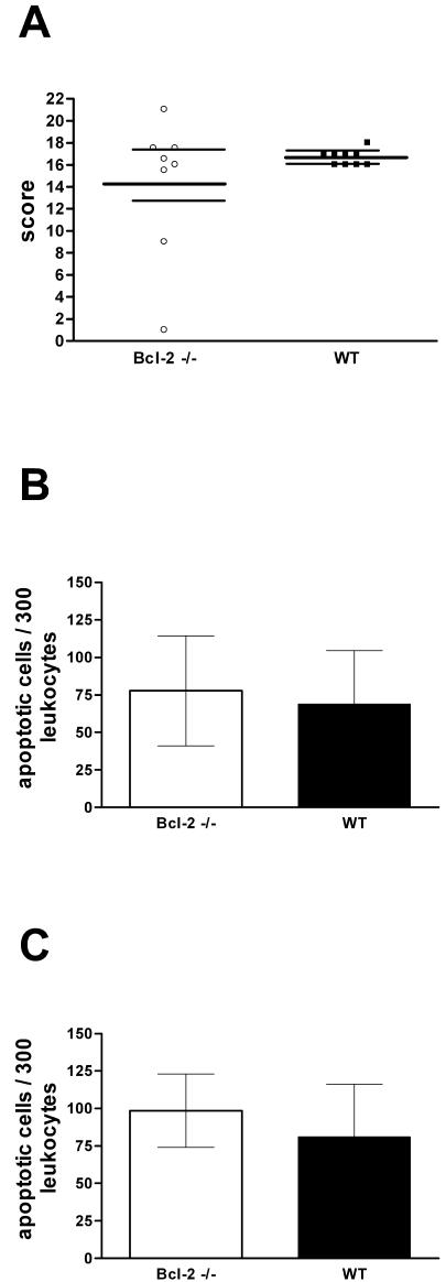
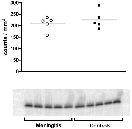
Necrotic and apoptotic neuronal cell death can be found in pneumococcal meningitis. We investigated the role of Bcl-2 as an antiapoptotic gene product in pneumococcal meningitis using Bcl-2 knockout (Bcl-2(-/-)) mice. By using a model of pneumococcal meningitis induced by intracerebral infection, Bcl-2-deficient mice and control littermates were assessed by clinical score and a tight rope test at 0, 12, 24, 32, and 36 h after infection. Then mice were sacrificed, the bacterial titers in blood, spleen, and cerebellar homogenates were determined, and the brain and spleen were evaluated histologically. The Bcl-2-deficient mice developed more severe clinical illness, and there were significant differences in the clinical score at 24, 32, and 36 h and in the tight rope test at 12 and 32 h. The bacterial titers in the blood were greater in Bcl-2-deficient mice than in the controls (7.46 +/- 1.93 log CFU/ml versus 5.16 +/- 0.96 log CFU/ml [mean +/- standard deviation]; P < 0.01). Neuronal damage was most prominent in the hippocampal formation, but there were no significant differences between groups. In situ tailing revealed only a few apoptotic neurons in the brain. In the spleen, however, there were significantly more apoptotic leukocytes in Bcl-2-deficient mice than in controls (5,148 +/- 3,406 leukocytes/mm2 versus 1,070 +/- 395 leukocytes/mm2; P < 0.005). Bcl-2 appears to counteract sepsis-induced apoptosis of splenic lymphocytes, thereby enhancing clearance of bacteria from the blood.
Figures

Similar articles
-
Matrix metalloproteinase-9 deficiency impairs host defense mechanisms against Streptococcus pneumoniae in a mouse model of bacterial meningitis.Neurosci Lett. 2003 Mar 6;338(3):201-4. doi: 10.1016/s0304-3940(02)01406-4. Neurosci Lett. 2003. PMID: 12581831
-
Effect of deficiency of tumor necrosis factor alpha or both of its receptors on Streptococcus pneumoniae central nervous system infection and peritonitis.Infect Immun. 2001 Nov;69(11):6881-6. doi: 10.1128/IAI.69.11.6881-6886.2001. Infect Immun. 2001. PMID: 11598062 Free PMC article.
-
Protective role of NF-kappaB1 (p50) in experimental pneumococcal meningitis.Eur J Pharmacol. 2004 Sep 13;498(1-3):315-8. doi: 10.1016/j.ejphar.2004.07.081. Eur J Pharmacol. 2004. PMID: 15364010
-
Experimental studies of pneumococcal meningitis.Dan Med Bull. 2010 Jan;57(1):B4119. Dan Med Bull. 2010. PMID: 20175949 Review.
-
Current concepts in the pathogenesis of meningitis caused by Streptococcus pneumoniae.Curr Opin Infect Dis. 2002 Jun;15(3):253-7. doi: 10.1097/00001432-200206000-00007. Curr Opin Infect Dis. 2002. PMID: 12015459 Review.
Cited by
-
Bacteremia causes hippocampal apoptosis in experimental pneumococcal meningitis.BMC Infect Dis. 2010 Jan 3;10:1. doi: 10.1186/1471-2334-10-1. BMC Infect Dis. 2010. PMID: 20044936 Free PMC article.
-
Regulation of the autophagic bcl-2/beclin 1 interaction.Cells. 2012 Jul 6;1(3):284-312. doi: 10.3390/cells1030284. Cells. 2012. PMID: 24710477 Free PMC article.
-
Chloroquine and inhibition of Toll-like receptor 9 protect from sepsis-induced acute kidney injury.Am J Physiol Renal Physiol. 2008 May;294(5):F1050-8. doi: 10.1152/ajprenal.00461.2007. Epub 2008 Feb 27. Am J Physiol Renal Physiol. 2008. PMID: 18305095 Free PMC article.
-
Nucleation of platelets with blood-borne pathogens on Kupffer cells precedes other innate immunity and contributes to bacterial clearance.Nat Immunol. 2013 Aug;14(8):785-92. doi: 10.1038/ni.2631. Epub 2013 Jun 16. Nat Immunol. 2013. PMID: 23770641 Free PMC article.
References
-
- Banasiak, K. J., T. Cronin, and G. G. Haddad. 1999. Bcl-2 prolongs neuronal survival during hypoxia-induced apoptosis. Mol. Brain Res. 72:214-225. - PubMed
-
- Bogdan, I., S. L. Leib, M. Bergeron, L. Chow, and M. G. Täuber. 1997. Tumor necrosis factor-alpha contributes to apoptosis in hippocampal neurons during experimental group B streptococcal meningitis. J. Infect. Dis. 176:693-697. - PubMed
-
- Bohr, V., O. B. Paulson, and N. Rasmussen. 1984. Pneumococcal meningitis. Late neurologic sequelae and features of prognostic impact. Arch. Neurol. 41:1045-1049. - PubMed
-
- Böttcher, T., J. Gerber, A. Wellmer, A. V. Smirnov, F. Fakhrjanali, E. Mix, J. Pilz, U. K. Zettl, and R. Nau. 2000. Rifampin reduces production of reactive oxygen species of CSF phagocytes and hippocampal neuronal apoptosis in experimental Streptococcus pneumoniae meningitis. J. Infect. Dis. 181:2095-2098. - PubMed
-
- Braun, J. S., R. Novak, H. K. Herzog, S. M. Bodner, J. L. Cleveland, and E. I. Tuomanen. 1999. Neuroprotection by a caspase inhibitor in acute bacterial meningitis. Nat. Med. 5:298-302. - PubMed
Publication types
MeSH terms
Substances
LinkOut - more resources
Full Text Sources
Other Literature Sources
Medical
